BACKGROUND AND AIMS
Eruptive vellus hair cysts (EVHC) are rare, benign follicular anomalies, typically presenting as multiple small papules that may be asymptomatic or pruritic.1 Their clinical appearance can mimic acneiform, infectious, or granulomatous dermatoses, often leading to diagnostic delays and under-recognition.2 The authors aim to report a case of EVHC in a young woman with Fitzpatrick skin phototype VI, to emphasise the importance of clinicopathological correlation in persistent papular eruptions.
MATERIALS AND METHODS
An otherwise healthy 29-year-old woman with Fitzpatrick skin phototype VI was referred to the Dermatology Department at Hospital Santa Maria, Unidade Local de Saúde Santa Maria, Lisbon, Portugal, for evaluation of lesions localised to the abdomen that had been present for approximately 1 year. The lesions consisted of multiple firm, erythematous-to-brownish, 2–4 mm-sized, domed papules located on the intermammary and upper abdominal regions. Dermoscopy consisted of a central round, hypopigmented structure with a subtle pinkish hue, surrounded by a light-brown halo and fine, peripheral scale. The lesions were associated with mild pruritus and occasional drainage of yellowish material. No systemic involvement or family history of similar lesions was identified. Previous treatment with topical fusidic acid and betamethasone yielded no improvement. A skin biopsy was performed for diagnostic clarification.
RESULTS
Histopathological examination showed a mid-dermal cyst lined by stratified squamous epithelium with a preserved granular layer, containing laminated keratin and multiple transversely and obliquely cut vellus hair shafts. The synchronous emergence of multiple lesions and their distribution supported the diagnosis of EVHCs (Figure 1). Given the benign nature of the condition and the absence of significant symptoms, conservative management with regular follow-up was proposed.
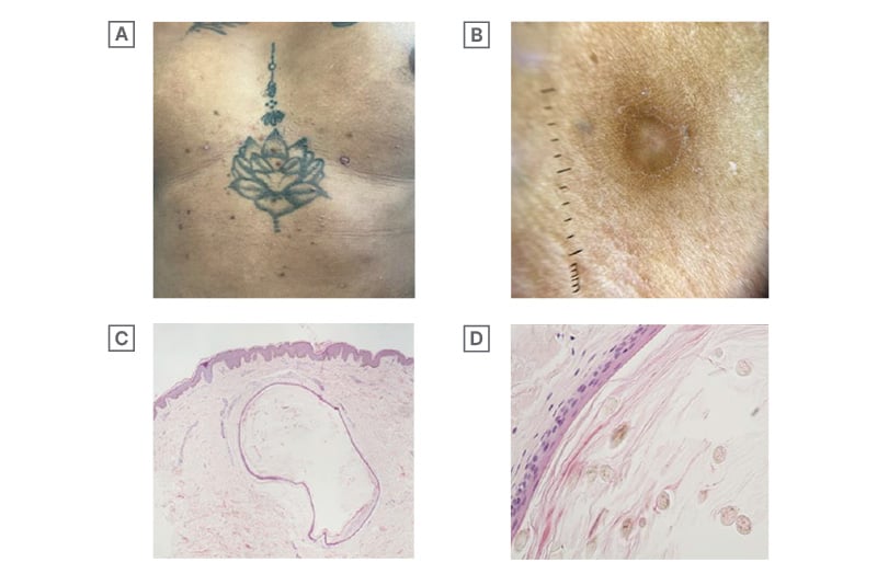
Figure 1: Eruptive vellus hair cysts.
A) Clinical image: multiple dome-shaped, pink-to-brownish-coloured papules on the abdominal region. B) Dermoscopic image: central hypopigmented area with a pinkish hue, surrounded by a brown halo and fine scaling (DermLite DL4; polarised mode; 10x magnification). C) and D) Histopathology features: mid-dermal cyst cavity containing laminated keratinous material and multiple vellus hair shafts (C: H&E, 40x magnification; D: 400x magnification).
DermLite DL4: DermLite, Aliso Viejo, California, USA; H&E: haematoxylin and eosin.
CONCLUSION
EVHCs are thought to arise froma developmental abnormality involving infundibular blockage, follicular dilatation, and hair bulb atrophy, with K17 gene mutations implicated in familial autosomal-dominant cases.3 This case underlines the relevance of considering EVHC in the differential diagnosis of chronic papular eruptions, particularly in patients with darker skin phototypes, where pigmentary changes may obscure classical features. Histopathological confirmation is keyto avoiding misdiagnosis andunnecessary treatments.