LARGE spot picosecond lasers potentially improve dermal pigment clearance in Asian skin while reducing perilesionaltissue exposure.
Large Spot Picosecond Laser Treatment Depends on Target Depth
A theoretical analysis evaluated how spot size and wavelength influence efficacy and safety for picosecond laser treatment of pigmented lesions in Asian skin. Researchers used a melanosome disruption threshold fluence model to estimate the fluence needed to disrupt melanosomes with 532, 730, 755, 785, and 1,064 nm picosecond lasers. To anticipate complication risk, they also simulated energy deposition in surrounding tissue, focusing on large spot irradiation, defined as spot sizes greater than 4 mm.
At 532 nm, spot size had minimal impact on the fluence required to disrupt epidermal melanosomes, and it produced little change in collateral energy deposition. Within the model assumptions, this suggests that both smaller and larger spot sizes could be viable for epidermal lesions at 532 nm, provided clinical endpoints and safety remain appropriate.
Near Infrared Wavelengths Shift the Spot Size Tradeoff
For wavelengths from 730 to 1,064 nm, the optimal spot size depended on lesion depth. When the target was epidermal melanosomes, smaller spot sizes concentrated energy in superficial layers, limiting exposure in deeper tissue. This depth focused deposition may be relevant when treating epidermal pigmentation where excessive penetration could increase unwanted heating in non-target structures.
When the target was dermal melanosomes, larger spot sizes reduced the required fluence by 39% to 65% and lowered collateral energy deposition in the simulations. Larger spots also decreased the variation in required fluence across lesion depths, potentially supporting more consistent parameter selection when depth is uncertain.
Early Clinical Feasibility at 1,064 nm
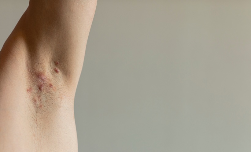
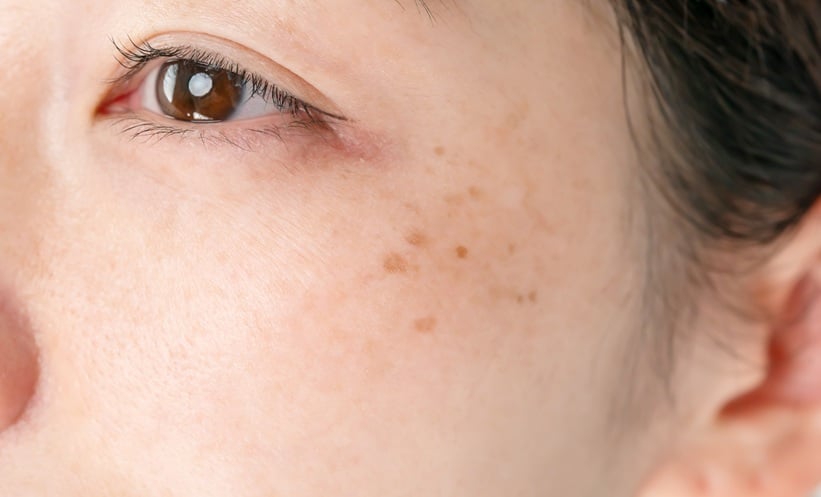
To validate feasibility, the authors reported a small clinical case series using a 1064 nm picosecond laser with a large spot size. Two dermal pigmented lesions, an ectopic Mongolian spot and a nevus of Ota, achieved good clearance without adverse events. The investigators noted that achieving sufficient efficacy with large spots may require higher energy output from laser devices, highlighting a practical consideration for device specifications and future studies.
Overall, the findings offer depth guided strategies for large spot picosecond laser treatment in Asian skin, aligning wavelength and spot size with the intended target layer to maintain melanosome disruption while reducing unintended energy deposition.
Reference: Shimojo Y et al. Theoretical Analysis of Large-Spot Picosecond Laser Treatment for Pigmented Lesions in Asian Skin. Lasers Surg Med. 2025;doi:10.1002/lsm.70086.