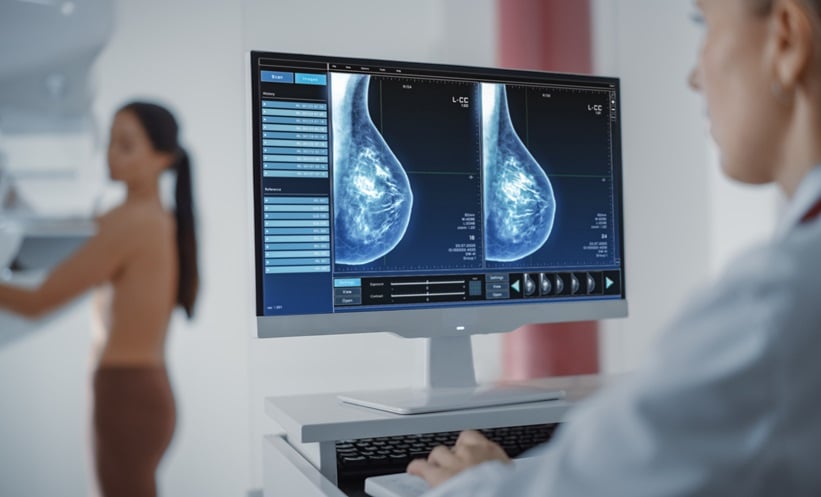
A NEW artificial intelligence model inspired by the work of human pathologists shows dramatically improves how breast cancer tissue is analysed, potentially reducing diagnostic delays and improving survival.
Digital Diagnosis Advances Through Breast Cancer Tissue Research
The global shortage of trained pathologists has placed significant pressure on timely cancer detection, particularly in regions where access to specialised care is limited. This has made accurate interpretation of breast cancer tissue samples increasingly challenging. Researchers at the University of Maine set out to address this gap by developing a system that can mimic a clinician’s visual workflow, offering a digital tool that strengthens diagnosis while maintaining clinical oversight. Their AI model, named CGS-Net, is designed to learn from both the microscopic details and the broader context of tissue slides, capturing the complexity of breast cancer tissue with greater precision than existing systems.
Dual-Encoder System Shows Superior Performance
To evaluate the model’s capabilities, the team trained CGS-Net using 383 digitised whole-slide images of lymph node tissue. The dual-encoder architecture was structured to imitate how pathologists analyse breast cancer tissue by switching between zoomed-out and highly magnified views. One encoder captured cell-level detail, while the second interpreted surrounding architecture. Each view shared the same centre pixel, ensuring precise alignment. The system used cross-attention features informed by a unique weight initialisation strategy. In tests, CGS-Net consistently outperformed single-input models, with an improvement in AUC ranging from 0.31 to 0.92 percent and an increase in cancer Dice score ranging from 4.09 percent to 6.81 percent. These findings signal meaningful progress in digital cancer detection and highlight the value of contextual image analysis.
Clinical Implications and Future Development
The study points toward a future in which AI-enhanced digital pathology supports faster, more reliable interpretation without replacing human judgement. For clinical practice, integrating tools like CGS-Net could streamline workflows, reduce diagnostic variability and improve the detection of early malignancy in breast cancer tissue. The researchers intend to expand the model to multiclass segmentation, incorporate additional magnification levels and adapt the architecture to other cancer types.
Reference
Juybari J et al. Context-guided segmentation for histopathologic cancer segmentation. Sci Rep. 2025;15:5404.