A SIMPLE geometric formula has shown promise in how clinicians assess carotid plaque volume, potentially offering a more accurate way to track atherosclerotic burden using routine ultrasound imaging.
Researchers reported that a hemi-ellipsoid formula, applied to standard carotid duplex ultrasonography images, enabled logical and reliable estimation of three-dimensional carotid plaque volume. This approach outperformed traditional one- and two-dimensional stenosis measures, which are commonly used in clinical practice but may underestimate disease severity.
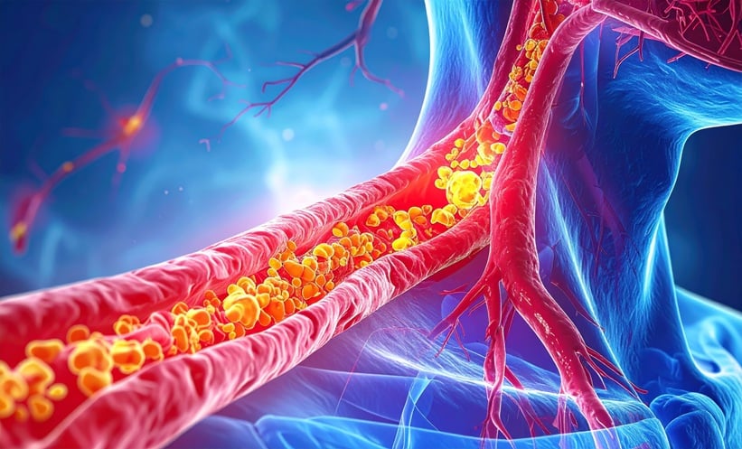
Carotid atherosclerosis is a major contributor to ischaemic stroke and cardiovascular disease worldwide. Current assessment typically relies on percentage diameter stenosis or area stenosis, which focus on luminal narrowing rather than total plaque burden. However, plaques develop and progress in three dimensions, meaning that volumetric assessment may better reflect true disease severity and risk.
Carotid Plaque Volume as a Better Disease Marker
In the study, investigators measured circumferential length, sagittal length, and mean luminally protruding thickness from axial and sagittal ultrasound images. These parameters were then entered into a hemi-ellipsoid volume formula to calculate carotid plaque volume. The authors logically validated that this method could be applied to plaques with smooth or irregular contours, as well as those involving part or all of the arterial lumen.
The researchers found that carotid plaque volume derived using the hemi-ellipsoid approach provided a more sensitive tool for evaluating plaque severity and monitoring progression over time compared with diameter or area stenosis alone. This suggests that volumetric assessment may better capture changes in atherosclerotic burden during follow-up.
Clinical Implications
For clinicians, carotid plaque volume measurement could address an important gap in vascular imaging. Carotid duplex ultrasonography is widely available, non-invasive, and inexpensive, making the adoption of a simple mathematical formula potentially feasible in routine care. A clearer picture of plaque burden may help refine risk stratification for patients with carotid artery disease.
The authors noted several limitations. Carotid duplex ultrasonography could not reliably assess plaque calcification, and standardised criteria for classifying plaque volume severity are lacking, while further studies are needed to confirm the applicability of the hemi-ellipsoid formula to highly irregular or ulcerated plaques.
However, this method could support more precise monitoring of atherosclerosis progression and treatment response, moving carotid imaging beyond stenosis towards a fuller assessment of vascular disease burden.
Reference
Kim J et al. Hemi-ellipsoid formula enables accurate assessment of carotid plaque volume and atherosclerotic burden. Sci Rep. 2026; DOI:10.1038/s41598-026-35182-5.