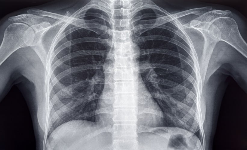
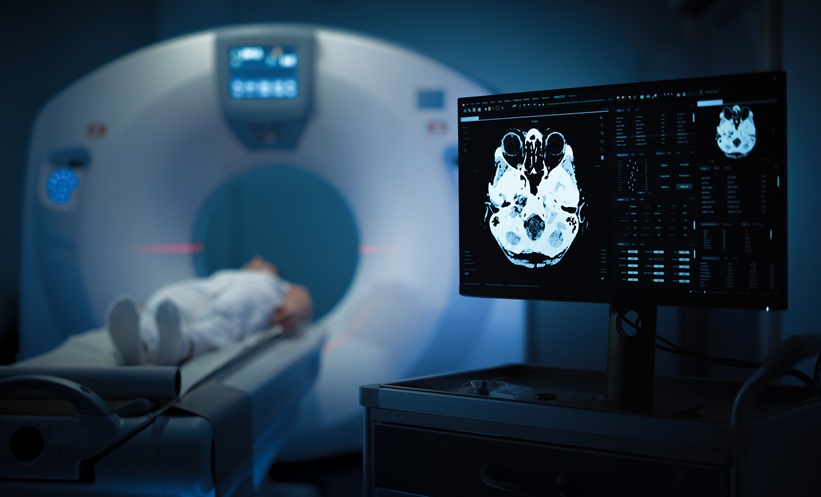
AI is increasingly being used to enhance diagnostic accuracy and streamline clinical workflows. One emerging application is the use of AI to detect osteoporosis and osteopenia from low-dose chest CT scans, which are commonly performed for other clinical reasons. A recent study aimed to systematically assess the accuracy of AI-based vertebrae segmentation and osteoporosis screening using such imaging. A key finding is that AI demonstrated excellent diagnostic performance, with pooled sensitivity and specificity values exceeding 86%.
A systematic review and meta-analysis was conducted in accordance with PRISMA-DTA guidelines. The authors searched PubMed, Scopus, Web of Science, and the Cochrane Library up to 13 December 2024. Studies evaluating AI models for vertebrae segmentation and osteoporosis detection from low-dose chest CT scans were included. Methodological quality was assessed using a modified QUADAS-2 tool. Meta-analytical techniques were employed to calculate pooled diagnostic accuracy statistics, and meta-regression and subgroup analyses explored potential sources of heterogeneity. Publication bias was assessed using the Egger test and funnel plots.
Eight studies met the inclusion criteria. For vertebrae segmentation, the pooled Dice similarity coefficient was 0.92 (95% CI 0.88–0.97), indicating high segmentation accuracy. For identifying individuals with abnormal bone mineral density (osteoporosis or osteopenia), pooled sensitivity was 0.90 (95% CI 0.88–0.91) and specificity was 0.90 (95% CI 0.88–0.91), with a summary receiver operating characteristic (SROC) of 0.9653. For detecting osteoporosis alone, pooled sensitivity was 0.86 (95% CI 0.82–0.89) and specificity was 0.93 (95% CI 0.92–0.94), with a SROC of 0.9676. Subgroup analyses identified significant heterogeneity based on dataset type, radiologist involvement in annotations, use of radiomics, and spinal region included.
This study demonstrates that AI models using low-dose chest CT images can accurately detect vertebral changes indicative of low bone mineral density. Clinically, this opens opportunities for opportunistic osteoporosis screening during routine imaging, potentially identifying at-risk patients earlier and more efficiently. However, the findings are limited by the small number of included studies and the reliance on retrospective data. Future prospective, multi-centre studies using diverse datasets are essential to validate these results and support clinical integration.
Reference
Ya’nan et al. Accuracy of Low-Dose Chest CT-Based Artificial Intelligence Models in Osteoporosis Detection: A Systematic Review and Meta-analysis. Calcif Tissue Int. 2025;DOI: 10.1007/s00223-025-01377-7.


