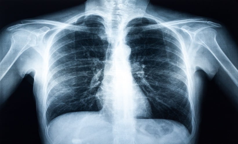
A novel form of chest X-ray imaging could help clinicians detect pneumothorax more quickly and with greater confidence, according to a prospective study evaluating dark-field chest radiography. Pneumothorax, caused by air leaking into the space around the lung, can be life-threatening if not identified promptly, particularly in emergency and critical care settings.
New radiography technique put to test
The study, conducted between March 2022 and September 2023, assessed whether adding dark-field imaging to conventional chest radiography could improve diagnostic performance. Dark-field radiography captures additional information about lung microstructure that is not visible on standard attenuation-based X-rays, potentially highlighting subtle abnormalities associated with air leakage.
Researchers examined 100 participants with a median age of 62 years, including 36 patients with clinically confirmed pneumothorax and 64 healthy controls. All participants underwent chest imaging using a prototype system capable of acquiring both conventional and dark-field images simultaneously, without requiring an additional scan.
Five radiologists, blinded to clinical information, independently reviewed the images. They first assessed standard chest radiographs alone and, after a four-week interval to minimise recall bias, reviewed images with a dark-field overlay. Sensitivity, specificity, accuracy, reading time and diagnostic confidence were then compared between the two approaches.
Faster reads with greater confidence
The results showed that adding dark-field overlays led to a small, non-significant increase in sensitivity for pneumothorax detection, rising from 84.2% with standard radiography to 87.4% with dark-field imaging. Specificity remained almost identical between the two methods, at around 97.5%, indicating that diagnostic accuracy was maintained.
The most striking improvement was seen in efficiency. Median reading time fell sharply from 30.8 seconds for conventional images to just 10.3 seconds when dark-field overlays were used, a reduction of nearly two-thirds. This difference was statistically significant and consistent across all readers.
Diagnostic confidence also improved. On a five-point scale, the median confidence score increased from 3 to 4 when radiologists assessed dark-field images, suggesting that clinicians felt more assured in their interpretations.
Clinical implications
The authors conclude that integrating dark-field radiography into routine chest X-ray assessment could offer meaningful clinical benefits. By substantially reducing reading time and improving confidence without compromising sensitivity or specificity, the technique may support faster decision-making, particularly in high-pressure environments such as emergency departments.
While larger studies are needed before widespread adoption, the findings highlight the potential of dark-field imaging as a valuable enhancement to conventional chest radiography for pneumothorax detection.
Reference
Gassert FT et al. Dark-field chest radiography for pneumothorax detection: a prospective study. Radiology: Cardiothoracic Imaging. 2025;7(6):e240560.