A novel imaging-based approach could simplify and improve radiation dose estimation in computed tomography, according to researchers who report strong performance across multiple adult scan regions. The technique aims to overcome practical barriers that have limited wider clinical use of size specific dose estimates.
Limits Of Current Dose Metrics
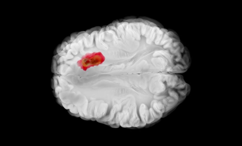
Computed tomography is one of the most widely used diagnostic imaging tools, but accurately estimating patient radiation dose remains challenging because of wide variation in body size and anatomy. Conventional dose metrics such as CT dose index volume and dose length product do not adequately account for patient specific factors.
Size specific dose estimates offer a more personalised assessment by incorporating the water equivalent diameter, a measure that reflects patient attenuation characteristics. However, calculating the water equivalent diameter for every image slice is time consuming and often impractical in routine clinical workflows. As a result, simplified approaches such as centre slice-based estimates are sometimes used, although these may reduce accuracy.
Slice Averaged Image Approach
To address this challenge, investigators developed a new method called the size specific dose estimate slice averaged image. Instead of calculating water equivalent diameter on a slice-by-slice basis, the method generates a single representative image by averaging computed tomography values across all slices in a scan. This averaged image is then used to calculate water equivalent diameter, capturing overall anatomical variability while substantially reducing computational effort.
The researchers retrospectively analysed computed tomography data from 282 adult patients. Three scan regions were included: chest, abdomen pelvis, and combined chest abdomen pelvis. For each examination, dose estimates derived using the new method were compared with centre slice-based size specific dose estimates and with a reference mean size specific dose estimate calculated across all slices.
Strong Agreement With Reference Standard
Across all scan regions, the slice averaged image approach showed stronger agreement with the reference mean size specific dose estimate than the centre slice method. Regression analysis demonstrated high coefficients of determination, with R² values reaching as high as 0.991. The new method also produced lower root mean square error values, indicating improved accuracy in dose prediction.
Reference
Dendo Y et al. A practical slice averaged image method for precise CT size specific dose estimates. Scientific Reports. 2025;15:42870.