Routine imaging used to stage prostate cancer may also offer a valuable opportunity to identify osteoporosis and fracture risk, according to new research examining men receiving androgen deprivation therapy.
Androgen deprivation therapy is a cornerstone of prostate cancer treatment but is well known to accelerate bone loss and increase the risk of fragility fractures. Despite this, baseline bone mineral density assessment is rarely performed in clinical practice. Researchers investigated whether information already captured during standard cancer imaging could be repurposed to improve bone health screening in this high-risk group.
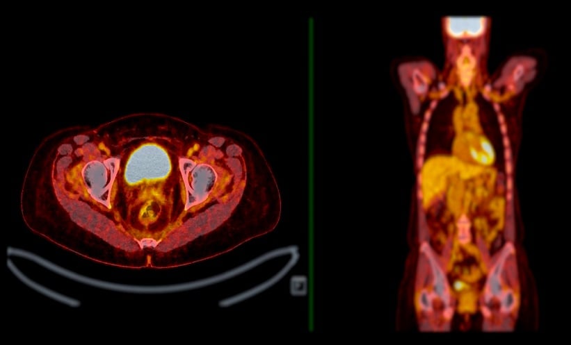
Comparing PET CT And Diagnostic CT
The retrospective single-centre study included 81 men with prostate cancer who underwent both 18F-fluorocholine PET/CT and stand-alone diagnostic CT within a 6 month period. Investigators measured trabecular attenuation in Hounsfield units at the L1 to L3 vertebral levels on both types of scans, using attenuation as a surrogate marker of bone mineral density.
Agreement between trabecular attenuation measurements from PET/CT and diagnostic CT was good across all vertebral levels, with intraclass correlation coefficients of 0.72 at L1, 0.70 at L2 and 0.67 at L3. Mean trabecular attenuation values measured on PET/CT were 121 HU at L1, 122 HU at L2 and 118 HU at L3, demonstrating close comparability with stand-alone CT measurements.
These findings indicate that the low-dose CT component of PET/CT, already performed as part of routine prostate cancer care, can reliably provide information relevant to bone health.
Links With Therapy Duration And Fractures
Longer exposure to androgen deprivation therapy was significantly associated with lower trabecular attenuation, with a moderate negative correlation observed between attenuation values and therapy duration. This supports existing evidence that prolonged hormonal treatment contributes to progressive bone loss.
Vertebral fractures were identified in 18.5% of patients and were strongly associated with lower trabecular attenuation. Patients with fractures had a mean attenuation of 87.8 HU compared with 127.5 HU in those without fractures, highlighting the clinical relevance of attenuation measurements for fracture risk assessment.
Implications For Clinical Practice
The study suggests that opportunistic assessment of bone mineral density using PET/CT could help address a major gap in osteoporosis screening among men with prostate cancer. By integrating trabecular attenuation measurements into routine imaging workflows, clinicians may be able to identify patients at high fracture risk without additional scans, radiation exposure or cost.
Researchers conclude that this approach could support earlier intervention to protect bone health, reduce fracture rates and improve long-term outcomes for men receiving androgen deprivation therapy.
Reference
Dauchez A et al. Opportunistic screening for osteoporosis and vertebral fracture using CT attenuation from 18F-fluorocholine PET/CT in patients with prostate cancer. RMD Open. 2025;11:e006231.