INTRODUCTION
BK virus, also known as Polyomavirus hominis 1, is a common virus that infects about 60–90% of the general population during childhood and then remains latent in the kidneys. Reactivation can occur in the setting of immune suppression. Infection with BK virus causing hemorrhagic cystitis has been well documented in patients following hematopoietic stem cell transplantation (HSCT), kidney transplants, and HIV infections.1 However, BK virus causing pneumonia has rarely been reported in the literature. Here the authors present a case of a patient with HSCT complicated by hemorrhagic cystitis and respiratory failure, with concern for BK viral pneumonia.
CASE
A 22-year-old male with a history of B cell acute lymphoblastic leukemia and HSCT was presented to the hospital for graft-versus-host disease management. After poor response to initial management, it was recommended to repeat a liver biopsy, which was complicated by right hemothorax and drained by interventional radiology intraoperatively. His course was then complicated by re-accumulation of the hemothorax with respiratory distress, so he was admitted to the pediatric ICU. His respiratory failure progressed, with new multifocal opacities observed on chest X-ray, and requiring intubation. The chest tube continued to drain copious amounts of blood, so a CT with angiography was obtained to rule out a vascular injury causing hemothorax. The CT revealed multifocal ground-glass opacities and right lower lobe consolidation, representing pulmonary hemorrhage or infection (Figure 1). Due to his immune deficient status, a bronchoscopy with bronchoalveolar lavage (BAL) was obtained to investigate for opportunistic infections.
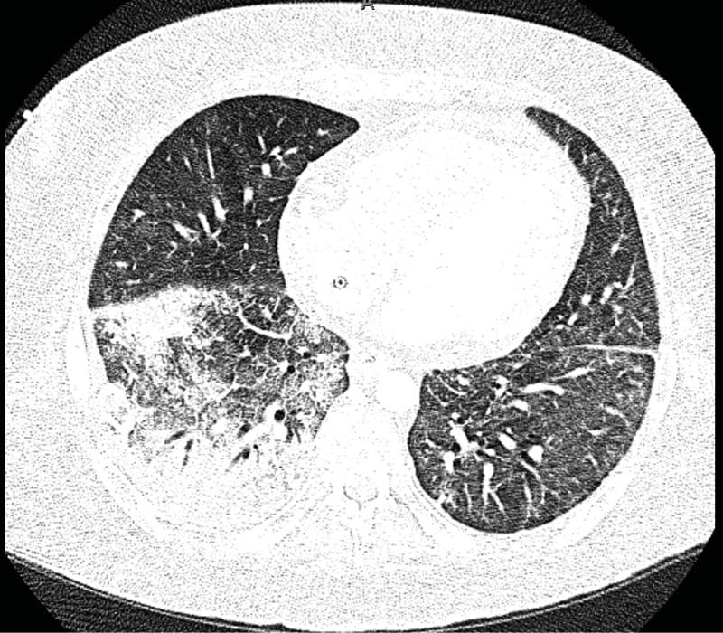
Figure 1: CT with angiography of the chest showing multifocal ground-glass opacities and consolidation in the right lower lobe, that may represent hemorrhage, infection, and/or aspiration.
There was no evidence of vascular injury causing hemothorax or pulmonary hemorrhage.
The patient had a history of BK virus positive urine and developed new hemorrhagic cystitis during his pediatric ICU stay. His BK viral count in the urine was >500,000,000 copies/mL, and he also had significant viremia, prompting BK virus testing in the BAL sample. BAL studies were significant only for a BK viral count of 1,700 copies/mL. On cytology, the sample was markedly hemorrhagic, with no evidence of fungus on silver stain and 2–4% lipid-laden macrophages. Due to the severity of his graft-versus-host disease, his medical team determined that he was not a candidate for the typical therapy for BK virus, which is the removal of immunosuppressive medications. Unfortunately, the patient continued to decompensate, and he passed away from multiple organ failure.2
DISCUSSION
Per literature review, there have been very few cases reported of suspected BK viral pneumonia, with only three other cases reported where infection was confirmed with BAL.1,3-7 The typical treatment for BK virus nephropathy typically includes withdrawal of immunosuppression, which was not possible in this patient. Other treatments that have been attempted for both nephropathy and pneumonia include cidofovir, leflunomide, quinolone antibiotics and intravenous immunoglobin; however, the treatment is not standardized and was used with varying success.1-7 Due to the rarity of cases, it is unclear whether the finding of BK virus in the BAL proves the causality of his pneumonia, or whether it is only a marker of the degree of his immune suppression. In this case, no other infectious etiology was found to explain his clinical symptoms, leading the authors to believe that the BAL was indicative of infection causing disease. Collectively, these cases suggest that the finding of BK virus in the lungs is associated with a high mortality rate.