NOVEL cartilage imaging methodologies are sharpening identification of joint damage, and monitoring response in rheumatic diseases.
MRI Still Leads Cartilage Imaging Techniques
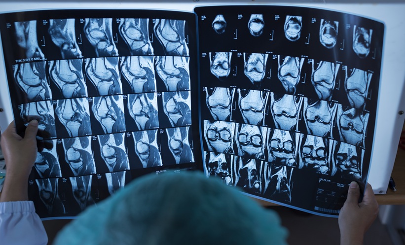
Non-invasive imaging remains central to diagnosing and tracking cartilage pathology in rheumatology, particularly where cartilage damage is clinically meaningful but difficult to reverse. In this review, MRI is positioned as the reference standard for directly visualizing articular cartilage, with the ability to assess both morphology and composition. Higher magnetic field strengths can support finer spatial resolution and faster acquisitions, while semi-quantitative scoring helps characterize the extent and depth of cartilage surface damage and can incorporate related joint structures, including subchondral bone and menisci.
Cartilage Imaging Techniques Detect Matrix Change Earlier
A key shift described is the growing clinical relevance of compositional MRI approaches that identify matrix alterations before overt surface breakdown. Techniques highlighted include T2 mapping, T1ρ, delayed gadolinium-enhanced MRI of cartilage, sodium imaging, diffusion imaging, and ultra-short echo-time imaging. These approaches can probe cartilage ultrastructure and may function as biomarkers of early degeneration, complementing quantitative cartilage morphometry. Morphometry relies on segmentation of cartilage tissue and computation of three-dimensional measures, and is described as highly sensitive to change over time, supporting longitudinal monitoring.
CT Arthrography and Emerging Modalities
When MRI is contra-indicated, CT arthrography is presented as a valuable alternative despite radiation exposure, with strong performance for detecting and evaluating cartilage surface lesions. The review also notes expanding, but still limited, roles for ultrasonography and PET to provide additional functional insight into cartilage and adjacent tissue processes.
Artificial Intelligence as an Imaging Catalyst
The authors anticipate artificial intelligence will increasingly reshape cartilage evaluation by accelerating MRI acquisition, enabling automated segmentation for precise cartilage quantification, and improving interpretive efficiency. As adoption grows, advanced cartilage imaging will likely support more individualized management of cartilage pathology across rheumatic disease.
Reference: Guermazi A et al. Advances in cartilage imaging techniques. Nat Rev Rheumatol. 2026;doi:10.1038/s41584-026-01353-x.