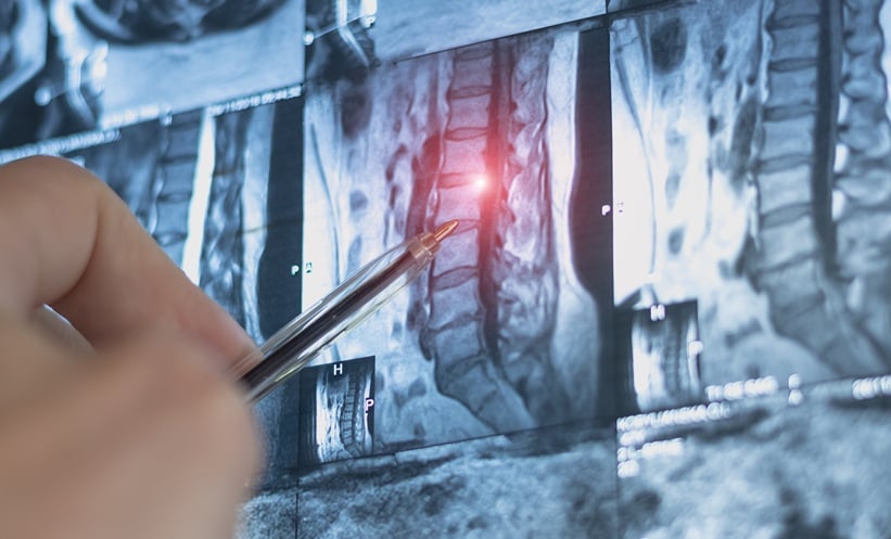
IN PATIENTS with early axial spondyloarthritis (axSpA), subtle but significant differences in structural spinal lesions are detectable on MRI but not on conventional radiography (CR), according to 2-year findings from the SPACE cohort presented at EULAR 2025.
Researchers compared structural progression in patients with chronic back pain, both with and without a diagnosis of axSpA, using both imaging modalities. While CR showed minimal progression over two years for both groups, MRI revealed a clear increase in fat lesions among patients with axSpA, but not in those with non-axSpA chronic back pain.
A total of 318 patients (214 axSpA, 108 non-axSpA) underwent CR and 351 (242 axSpA, 109 non-axSpA) had MRI data available for analysis. Over the two-year period, axSpA patients experienced a measurable increase in fat lesions (mean +0.5, p=0.01), with a greater proportion meeting cut-offs for ≥5 fat lesions or fat lesions combined with erosions (p=0.01). Notably, 10% of axSpA patients developed three or more new fat lesions versus just 1% in the non-axSpA group (p=0.001).
Despite the imaging differences, conventional scoring (mSASSS) on radiography remained largely unchanged for both groups, with negligible progression observed. The results suggest MRI fat lesions could serve as a more sensitive biomarker in early disease monitoring, offering potential utility in clinical decision-making.
Reference
Ayan G et al. Differences in structural lesions of the spine between patients with early axSpA and non-axSpA chronic backpain: 2-year results of the SPACE Cohort. Abstract: OP0310. EULAR Congress, 11-14th June, 2025.