Key Summary:
- Superficial dyskeratotic keratinocytes appeared in 12 of 16 skin biopsies.
- The finding occurred across several rash appearances, not only classic lesions.
- Larger prospective studies must confirm the diagnostic value of skin biopsy.
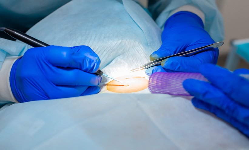
SKIN biopsy identified a potentially valuable diagnostic marker in adults with suspected adult-onset Still’s disease.
Adult-onset Still’s disease is a rare autoinflammatory condition that can present with fever, rash, joint symptoms, and elevated inflammatory markers. Diagnosis remains challenging because these manifestations overlap with sepsis, hemophagocytic lymphohistiocytosis, and other inflammatory syndromes. Patients may also lack the classic transient, salmon-colored rash recognized in current classification criteria.
Investigators retrospectively examined clinical records and skin biopsies from 16 adults with confirmed adult-onset Still’s disease who received care through two Canadian rare disease programs between 2016 and 2025. All patients had fever and arthralgia or polyarthritis. Four had catastrophic disease involving severe complications such as macrophage activation syndrome, disseminated intravascular coagulation, or myocarditis requiring intensive care.
Dyskeratotic or necrotic keratinocytes were identified in the superficial epidermis in 12 of 16 patients. These abnormal cells were found across diverse skin presentations, including macular and maculopapular lesions, erythematous rashes, and persistent pruritic eruptions.
The findings suggest that the histologic pattern may not be restricted to the persistent pruritic plaques with which it has traditionally been associated. Only one patient had a documented classic salmon-colored evanescent rash, highlighting the variability of cutaneous manifestations.
The researchers noted that superficial dyskeratotic keratinocytes can occur in several other conditions, including acute graft-versus-host disease, toxic shock syndrome, phototoxic reactions, and irritant contact dermatitis. However, these disorders generally arise in distinct clinical settings and can often be differentiated through patient history and examination.
Importantly, the finding has not been reported in several major adult-onset Still’s disease mimics, including sepsis, non–Still’s disease hemophagocytic lymphohistiocytosis, TAFRO syndrome, and VEXAS syndrome.
Skin biopsy is minimally invasive, widely available, and can often be interpreted within one working day. The investigators therefore proposed performing it early when adult-onset Still’s disease is suspected and a rash is present.
However, the study included only 16 patients and relied on retrospective records with variable descriptions of skin morphology. Larger prospective studies using standardized dermatologic examinations are needed before skin biopsy findings can be incorporated into diagnostic or classification criteria.
Reference
Chong DHY et al. Skin biopsy in adult-onset Still’s disease: A specific, rapid, and widely available diagnostic test. J Hum Immun. 2026;2(6):e20260088.
Featured Image: Kate on Adobe Stock.
Each article is made available under the terms of the Creative Commons Attribution-Non Commercial 4.0 License.