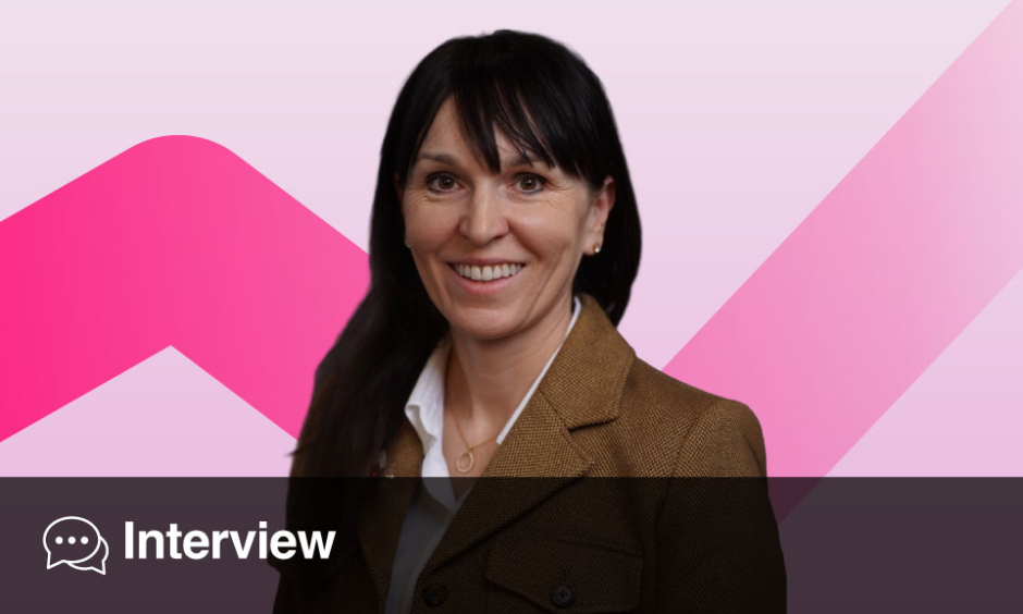
Gosia Wamil: Assistant Professor and Consultant Cardiologist, Mayo Clinic Healthcare, London, UK; Consultant Cardiologist, Great Western Hospital NHS Trust, Swindon, UK; Honorary Research Fellow, Deep Medicine, University of Oxford, UK
Citation: EMJ Cardiol. 2026;14[1]:119-125 https://doi.org/10.33590/emjcardiol/Q54YF209

You have spent more than two decades in cardiology and now lead multimodality cardiac imaging services in the NHS and at Mayo Clinic Healthcare in London, UK. Looking back, what were the pivotal moments that convinced you imaging could be more than a diagnostic tool and become a driver of clinical decision-making?
For much of my early training, imaging was presented as a confirmatory test: you formed a clinical impression and then ordered a scan to support it. What changed my thinking was seeing cardiovascular magnetic resonance (CMR) repeatedly overturn diagnoses that had seemed clinically convincing. A patient thought to have a non-specific cardiomyopathy might show a pattern of fibrosis that completely changes their diagnosis, prognosis, and management. Once you have seen imaging redirect the entire course of a patient’s care, you stop thinking of it as the final step and start seeing it as the point where many of the most important clinical decisions begin.
The cases that have stayed with me are those in which no single imaging modality provided the answer. A patient with unexplained left ventricular hypertrophy may initially appear to have hypertrophic cardiomyopathy (HCM) on echocardiography. However, when CMR demonstrates diffuse infiltration rather than sarcomeric disease, the diagnosis changes to cardiac amyloidosis, with entirely different treatment options and prognosis. Those are the moments that convinced me that multimodality imaging is not about performing more tests; it is about asking the next question that will change management.
That philosophy has shaped my career. Rather than thinking of echocardiography, CT, or CMR as separate disciplines, I have increasingly viewed them as complementary tools, each addressing a different clinical question. The goal is never to produce the most comprehensive imaging report; it is to provide the information that allows the multidisciplinary team to make the right decision for the patient. That, to me, is where imaging becomes far more than a diagnostic tool and becomes a driver of personalised care.
As both cardiovascular imaging and cardiomyopathy care become increasingly multidisciplinary, a shift formalised by the 2023 European Society of Cardiology (ESC) Guidelines on the management of cardiomyopathies, what lessons have you learned about collaboration across specialties, and how has this shaped your approach to patient care and research?
One of the biggest changes over the past decade has been recognising that no single specialist has all the answers in cardiomyopathy. These are complex diseases that sit at the intersection of imaging, genetics, electrophysiology, heart failure, and increasingly clinical pharmacology. The best decisions are rarely made in isolation.
Working in multidisciplinary teams has taught me that the value of imaging is not simply in producing an accurate report, but in providing the information that each specialist needs to answer a specific clinical question. The electrophysiologist may be focused on arrhythmic risk, the geneticist on whether a phenotype supports the interpretation of a genetic variant, and the heart failure specialist on whether imaging suggests a potentially treatable cause. The same scan can therefore mean very different things depending on the clinical context.
That is why I believe some of the most valuable work happens around the multidisciplinary meeting table. Reviewing images together, discussing uncertainty openly, and combining different perspectives almost always leads to better decisions than any one speciality could reach alone.
The same philosophy has shaped my research. The most interesting questions often arise at the boundaries between disciplines, where imaging meets genetics, heart failure, electrophysiology, or cardiometabolic medicine. Ultimately, multidisciplinary care is not about having more experts involved; it is about ensuring that every patient benefits from the combined expertise of the whole team. That, in my experience, is how imaging has its greatest impact.
Your expertise spans echocardiography, cardiac CT, and CMR at a time when cardiovascular imaging is becoming increasingly subspecialised. How have you navigated these rapidly evolving technologies while maintaining a patient-centred approach to care?
The challenge of modern cardiovascular imaging is not keeping up with technology; it is knowing when that technology genuinely benefits the patient. As imaging has become increasingly specialised, there is a risk of becoming an expert in a modality rather than an expert in solving clinical problems.
I have always enjoyed working across echocardiography, cardiac CT, and CMR because each offers a different perspective. Echocardiography excels at physiology and haemodynamics, CT provides unparalleled assessment of coronary anatomy and atherosclerosis, and CMR offers unique insights into myocardial tissue and disease mechanisms. The real value lies not in mastering each technique individually, but in understanding how they complement one another.
At the same time, a patient-centred approach sometimes means recognising that the best investigation is no further investigation at all. Every scan carries costs, potential risks, and the possibility of incidental findings that may create more uncertainty than clarity. One of the most important skills an experienced imager develops is knowing when enough information has been obtained to make a confident clinical decision.
Ultimately, I see technology as an enabler rather than the destination. Patients rarely remember which imaging modality they underwent; they remember whether they received a clear diagnosis, an appropriate treatment plan, and confidence that the decisions being made were the right ones. That perspective has helped me embrace new technologies while keeping the focus where it belongs: on the person rather than the scan.
Many clinicians still think of imaging modalities in silos. Throughout your career, you have championed a genuinely multimodal approach. Can you share an example where integrating several imaging techniques fundamentally changed a diagnosis, risk assessment, or management strategy?
One example that has stayed with me comes from a patient with longstanding Type 2 diabetes who presented with progressive exertional breathlessness despite a preserved ejection fraction. If we had relied on a single imaging modality, we would almost certainly have missed the underlying diagnosis.
Coronary CT angiography excluded significant coronary artery disease, allowing us to move beyond an ischaemic explanation. Echocardiography then demonstrated impaired global longitudinal strain with evidence of raised filling pressures, suggesting early myocardial dysfunction despite a normal ejection fraction. Finally, CMR revealed diffuse expansion of the extracellular volume without focal scar, consistent with diffuse interstitial fibrosis.
Taken together, these findings identified early diabetic cardiomyopathy, a diagnosis that no individual imaging test could have established with the same level of confidence. More importantly, it changed management. Rather than pursuing further investigations, we focused on aggressive cardiometabolic optimisation, including initiation of sodium-glucose cotransporter 2 (SGLT2) inhibition and intensive risk-factor modification.
For me, that is the essence of multi-modality imaging. It is not about performing more scans; it is about combining complementary information to answer questions that no single technique can answer alone. When imaging moves beyond describing anatomy and starts changing diagnosis, treatment, and prognosis, it becomes a true partner in clinical decision-making.
As a member of the European Association of Cardiovascular Imaging (EACVI) Scientific Committee, you have an unusual vantage point on developments across European cardiovascular imaging. Which emerging trends or research areas do you believe are currently under-appreciated, but have the potential to significantly influence practice in the coming years?
Three areas stand out to me. First, imaging is moving from diagnosis towards precision risk prediction. We already acquire vast amounts of imaging data, but the next step is integrating quantitative imaging biomarkers into clinical risk models that genuinely improve decision-making, rather than reporting them in isolation. The challenge is no longer acquiring better images but extracting clinically meaningful information.
Second, I believe imaging as a marker of treatment response is still under-appreciated. As disease-modifying therapies emerge for conditions such as HCM, amyloidosis, and heart failure, imaging will increasingly be used to detect early reverse remodelling and guide treatment escalation or de-escalation, rather than simply confirming a diagnosis.
Finally, the least glamorous but perhaps most important area is data standardisation. If we want imaging biomarkers to become part of routine care, they must be reproducible across vendors, centres, and healthcare systems. Harmonised acquisition protocols, structured reporting, and inter-operability are essential if we are to combine imaging with genomics, biomarkers, and electronic health records at scale.
From my perspective on the EACVI Scientific Committee, there is tremendous innovation across Europe. The opportunity now is to translate that innovation into robust evidence and practical workflows. Ultimately, the biggest advances over the next decade may not come from a new imaging modality, but from making the information we already acquire more quantitative, reproducible, and actionable for everyday clinical practice.
HCM has undergone a remarkable transformation, with new imaging techniques, risk-stratification tools, and therapeutic options now available. Which advances have had the greatest impact on patient care, and what do you consider the most pressing unanswered questions in the field today?
HCM is one of the few areas in cardiology where we’ve seen simultaneous advances in imaging, risk prediction, and disease-specific therapy. Together, they have fundamentally changed patient care.
From an imaging perspective, CMR has had the greatest impact. Beyond refining the diagnosis, late gadolinium enhancement has become central to sudden cardiac death risk assessment, particularly in patients whose conventional risk estimates sit in the grey zone. It has moved us away from relying solely on wall thickness and family history towards a more personalised understanding of arrhythmic risk.
Therapeutically, the introduction of cardiac myosin inhibitors represents a genuine paradigm shift. For the first time, we have treatments that target the underlying pathophysiology of obstructive HCM rather than simply alleviating symptoms. They have also changed the role of the imaging cardiologist, with serial echocardiography now guiding treatment initiation, dose titration, and long-term monitoring.
Despite this progress, important questions remain. Sudden cardiac death risk prediction is still imperfect, and we need more precise models that integrate imaging, genetics, and clinical data to better identify the patients who truly benefit from an implantable cardioverter defibrillator. We also need to understand whether myosin inhibitors alter the natural history of the disease by preventing fibrosis and adverse remodelling, rather than simply improving haemodynamics and symptoms.
Finally, I think the greatest unmet need is in non-obstructive HCM and genotype-positive, phenotype-negative individuals. These are often the most challenging conversations in the clinic because patients want to know not only what their diagnosis is today, but what their future is likely to look like. Helping us answer that question is where the next generation of imaging research should focus.
Cardiac MRI has become increasingly central to HCM assessment, particularly for tissue characterisation and risk stratification. How do you see its role evolving, and are we approaching the point at which imaging biomarkers could influence treatment decisions as much as traditional clinical parameters?
I think we’ve already crossed the point where CMR is no longer an adjunct; it is a core component of HCM assessment. Echocardiography remains the first-line investigation, but CMR provides information that simply cannot be obtained any other way, particularly around myocardial fibrosis and tissue characterisation. That information increasingly influences some of the most important decisions we make, especially around sudden cardiac death risk and implantable cardioverter defibrillator implantation.
Late gadolinium enhancement is the clearest example. We now have robust evidence that the extent of fibrosis provides incremental prognostic value beyond traditional clinical risk factors, particularly in patients who sit in the intermediate-risk category. Equally important, the absence of significant fibrosis can be reassuring and helps support a more conservative management strategy. In that sense, imaging biomarkers are already influencing treatment decisions alongside conventional clinical parameters.
Looking ahead, I think the next frontier is moving beyond replacement fibrosis towards earlier markers of disease activity. Native T1 mapping, extracellular volume quantification, and other quantitative CMR biomarkers may allow us to identify myocardial remodelling before irreversible scar develops. As disease-modifying therapies become more widely available, those biomarkers could help identify patients who would benefit from earlier intervention and provide objective measures of treatment response.
That said, imaging biomarkers should complement, not replace, clinical judgement. Their greatest value lies in integrating imaging with genetics, symptoms, rhythm assessment, and family history to deliver genuinely personalised care. Achieving that will require continued standardisation of quantitative CMR across centres, which I see as one of the key priorities for our field over the next decade.
AI is increasingly being integrated into cardiovascular imaging workflows. From your experience, where is AI already adding genuine clinical value, and where do you think expectations currently exceed reality?
AI is already adding genuine value, but perhaps not in the areas that attract the most headlines. Its greatest impact today is in automating repetitive, measurement-intensive tasks such as chamber quantification, ejection fraction, strain analysis, image segmentation, and quality control. These applications improve reproducibility, reduce reporting time, and allow clinicians to spend more time interpreting studies and discussing management with patients. In busy imaging departments, that consistency is a real advantage.
Where I think expectations currently exceed reality is in fully autonomous diagnosis and risk prediction. Many AI models perform extremely well in research datasets but are far less convincing when applied across different scanners, institutions, and patient populations. Before we rely on these tools for major clinical decisions, we need robust prospective validation demonstrating not just technical accuracy, but improved patient outcomes.
Looking ahead, I believe the greatest opportunity for AI is not replacing imaging specialists but helping us integrate increasingly complex information. Modern cardiovascular imaging generates an enormous amount of data, and AI can help combine imaging with clinical variables, genomics, biomarkers, and electronic health records to deliver more personalised risk assessment and treatment recommendations.
Ultimately, the future of AI in cardiovascular imaging is unlikely to be about replacing human expertise. It will be about making clinicians more efficient, more consistent, and, most importantly, enabling better-informed decisions for individual patients.
Alongside your clinical and research roles, you are heavily involved in education and standard setting through the EACVI and the British Society of Echocardiography (BSE). What skills will the next generation of cardiac imagers and consultant cardiologists need in order to succeed in an increasingly technology-driven field?
Technology will undoubtedly change how we practise, but I don’t think it changes what makes an excellent cardiologist. The next generation will need to be comfortable working across imaging modalities and understand how to use AI and quantitative tools, but technology should support clinical judgement, not replace it.
The skill I value most is the ability to ask the right clinical question before choosing the imaging test. An outstanding imager is not the person who performs the most sophisticated scan, but the one who understands what information will change management and selects the right modality to answer that question.
Equally important is the ability to integrate information. Modern cardiologists need to bring together echocardiography, CT, CMR, genetics, biomarkers, and clinical history into a single, coherent assessment. Patients do not experience their disease in silos, and neither should we.
Finally, I hope we continue to emphasise communication and curiosity. Imaging reports should inform decisions, not simply describe findings, and we must be able to explain uncertainty to both colleagues and patients. At the same time, the field is evolving so rapidly that continuous learning is no longer optional, it is part of the job.
Through my work with the EACVI, one of my priorities has been to help develop training and standards that prepare cardiologists not just for today’s practice, but for the technologies and therapies they will be using 10 years from now. In my view, the best cardiac imagers of the future will combine technical excellence with clinical insight, intellectual curiosity, and a commitment to lifelong learning.
As someone who combines clinical practice, research, society leadership, and education across two health systems, what has been the most rewarding aspect of your career so far, and what continues to motivate you today?
What has been the most rewarding is seeing how imaging can genuinely change a patient’s journey. Sometimes that means making a difficult diagnosis that had previously been missed; sometimes it means giving someone reassurance that they do not need an invasive procedure or device. Those conversations stay with you far longer than any publication or presentation because you can see the immediate impact on the patient and their family.
Beyond individual patients, I have found enormous satisfaction in helping shape the field through research, education, and professional societies. Whether contributing to clinical guidelines, developing imaging standards through the EACVI and the BSE, or mentoring trainees, there is a multiplier effect. If you help one clinician make better decisions throughout their career, the benefit extends to thousands of patients.
What continues to motivate me is that cardiovascular imaging remains one of the most dynamic areas of medicine. We are entering an era where advanced imaging, genetics, AI, and novel therapies are converging to deliver increasingly personalised care. It is a privilege to be part of that transformation.
Above all, I remain driven by curiosity. Every patient teaches us something new, and every scientific advance raises another question. That combination of clinical practice, research, and education means that no 2 days are the same. If, over the course of my career, I can contribute to better evidence, better imaging, and better training for the next generation of cardiologists, I will feel I have made a meaningful difference.
Finally, if we were to reconvene 10 years from now, what major changes do you think we would see in the diagnosis and management of cardiomyopathies, and what role do you hope imaging will play in that future?
I think the biggest change will be that we stop thinking about cardiomyopathies as diseases defined by how the heart looks and instead classify them by their underlying biology. Imaging, genetics, biomarkers, and clinical data will be integrated to identify the specific disease mechanism in each patient, allowing us to move from a ‘one-size-fits-all’ approach to truly personalised care.
In that future, imaging will no longer be used simply to confirm a diagnosis. It will help identify disease at a much earlier stage, refine individual risk, guide treatment selection, and monitor response to therapy over time. As more disease-modifying treatments become available, imaging will increasingly act as a biomarker, allowing us to determine whether a therapy is genuinely altering the natural history of disease rather than simply improving symptoms.
I also hope that AI will have matured beyond today’s excitement. Rather than being viewed as a separate technology, it should become an invisible part of everyday practice, improving workflow, integrating complex data, and supporting clinical decisions, while leaving clinicians free to focus on the patient in front of them.
Ultimately, I hope that in 10 years’ time we will be diagnosing cardiomyopathies earlier, treating them more precisely, and preventing more cases of heart failure and sudden cardiac death than we do today. If imaging evolves from producing beautiful pictures to becoming a central tool for personalised decision-making throughout the patient’s journey, I think we will have achieved something truly transformative.