Abstract
Background: AI is rapidly reshaping cardiovascular medicine, particularly within imaging-intensive fields such as interventional cardiology. Advances in machine learning and deep learning have enabled automated analysis of complex cardiovascular datasets and opened new opportunities for decision support across the coronary care continuum.
Objectives: This review summarises current and emerging applications of AI in interventional cardiology, spanning pre-procedural imaging, intraprocedural guidance, outcome prediction, and future technological developments.
Key findings: In the pre-procedural setting, AI-enhanced coronary CT angiography, AI-derived fractional flow reserve, and automated plaque characterisation improve risk stratification and patient selection for invasive angiography. Within the catheterisation laboratory, AI applications include automated angiographic interpretation, intravascular ultrasound and optical coherence tomography analysis, lesion classification, optimisation of stent sizing and deployment, emerging roles in structural heart interventions, and periprocedural haemodynamic monitoring. Machine learning models are also increasingly used for risk stratification, predicting bleeding, contrast-induced nephropathy, and mortality after percutaneous coronary intervention. Despite these advances, important challenges remain, including regulatory oversight, algorithm validation, bias mitigation, data governance, clinician education, and unresolved liability issues.
Conclusion: AI is evolving from a diagnostic imaging adjunct to a comprehensive decision-support platform in interventional cardiology. Emerging technologies such as real-time AI-assisted catheterisation laboratory systems, multimodal data integration, digital twin modelling, and AI-guided robotic interventions may further transform coronary intervention workflows, positioning AI as a procedural co-pilot that augments rather than replaces physician expertise.
Key Points
1. Interventional cardiology is a dynamic specialty that is very well suited for AI integration, because the field relies on high-resolution imaging and a vast variety of quantifiable data points that can aid the development of predictive models to augment established clinical and procedural workflows.2. This is a narrative review that synthesises the current and emerging applications of AI across the full scope of interventional cardiology practice. From pre-procedural imaging and intraprocedural guidance to post-procedural outcome prediction, this review follows the sequential pathway of current and future AI integration in interventional cardiology.
3. The role of AI in interventional cardiology is evolving from an ‘imaging gatekeeper’ that merely refines patient selection to a ‘procedural co-pilot’ that augments physician decision-making and patient outcomes. The safe and effective adoption of this technology will require robust validation, bias mitigation, and data governance to maximise future utility.
INTRODUCTION
AI refers to computational systems capable of performing tasks that traditionally require human cognitive abilities, including pattern recognition, classification, prediction, and decision-making. In cardiovascular medicine, AI has rapidly evolved from early rule-based approaches towards data-driven models capable of extracting clinically meaningful information from large and complex datasets. Advances in machine learning and deep learning have enabled automated analysis of cardiovascular imaging, physiological signals, and clinical data, creating new opportunities for decision support across the continuum of coronary artery disease management.1-5
Interventional cardiology is particularly well-suited to AI integration because the specialty relies heavily on high-resolution imaging, structured procedural workflows, and quantifiable anatomical and physiological measurements. Coronary angiography, coronary CT angiography (CCTA), intravascular ultrasound (IVUS), optical coherence tomography (OCT), and physiological indices such as fractional flow reserve (FFR) generate large volumes of standardised digital data that are highly amenable to machine learning analysis.6,7 These characteristics provide an ideal environment for the development of computational tools capable of assisting clinicians in lesion assessment, procedural planning, and risk prediction.
Recent advances in deep learning-based imaging analysis have significantly expanded the role of non-invasive and invasive coronary imaging. Automated algorithms can now perform coronary artery segmentation, quantify plaque burden, and detect stenosis on CCTA with high reproducibility.8-10 Similar approaches are increasingly applied to invasive angiography and intravascular imaging to support lesion characterisation and procedural optimisation. In addition, computational models capable of deriving physiological metrics such as FFR from CCTA datasets illustrate how AI can bridge anatomical imaging with functional assessment, improving patient selection for invasive angiography and revascularisation.11-13
Beyond imaging interpretation, AI techniques are increasingly applied to procedural risk prediction and outcome modelling. Machine learning models that integrate clinical variables, imaging metrics, and procedural data have demonstrated the ability to estimate individualised risks of complications such as bleeding, contrast-induced kidney injury, and mortality following percutaneous coronary intervention (PCI). These predictive frameworks may support more personalised treatment decisions and improve resource utilisation in interventional cardiology.14,15
Several structural characteristics of interventional cardiology further facilitate the integration of AI into clinical practice. The field involves time-sensitive decision-making, standardised procedural workflows, and quantitative imaging endpoints that can serve as objective reference standards for algorithm training and validation. In addition, growing regulatory approval of AI-enabled imaging tools and increasing availability of large cardiovascular datasets have accelerated translational efforts in recent years.8,15
When discussing these ever-evolving tools and techniques, it is important to note that most of the applications discussed in this review are at varying stages of validation and real-world deployment. While some tools have gained regulatory clearance at the federal level and achieved multicentre validation, others remain experimental and confined to the research laboratories. Additionally, much of the current literature focuses on robust performance at high-volume academic or research-focused centres, but implementation in a community and under-resourced institutions might reveal unforeseen limitations.
Several existing reviews have addressed the implementation of AI in interventional cardiology but largely focus on isolated domains such as cardiovascular imaging or risk prediction, with limited integration across the full interventional cardiology workflow.16-18 This review follows the sequential patient pathway from pre-procedural imaging through intraprocedural guidance, structural and complex procedures, and outcome prediction within a single cohesive framework, rather than addressing one domain in isolation.
Taken together, these developments suggest that AI is evolving from a diagnostic imaging adjunct towards a broader decision-support framework across the interventional cardiology workflow. Rather than replacing physician expertise, AI technologies may enhance clinical judgement by improving image interpretation, supporting procedural planning, and enabling more precise risk stratification. These developments are progressively shifting the role of AI from a non-invasive imaging ‘gatekeeper’ that refines patient selection to a potential procedural ‘co-pilot’ capable of assisting clinicians during coronary interventions. The emerging role of AI across the continuum of coronary artery disease management, from non-invasive imaging to intraprocedural guidance and outcome prediction, is illustrated in Figure 1.
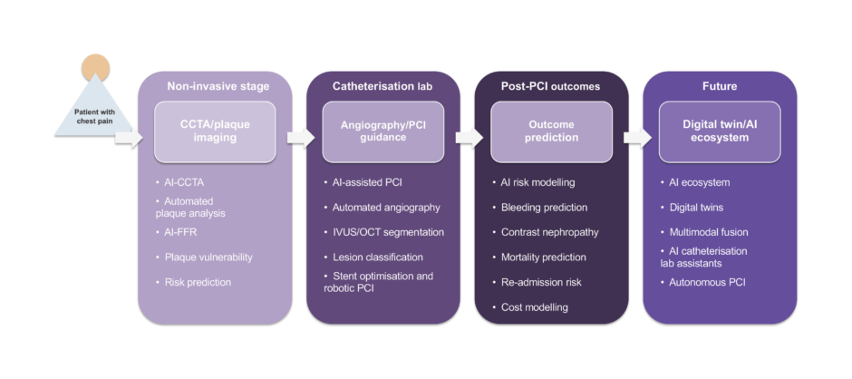
Figure 1: AI across the interventional cardiology workflow, from non-invasive imaging to procedural guiance.
CCTA: coronary CT angiography; FFR: fractional flow reserve; IVUS: intravascular ultrasound; OCT: optical coherence tomography; PCI: percutaneous coronary intervention.
AI BEFORE THE CATH LAB
Before a patient reaches the catheterisation laboratory, AI is increasingly influencing diagnostic pathways and procedural decision-making. Traditionally, progression from symptom evaluation to non-invasive imaging and invasive angiography has relied on clinician interpretation and population-based risk models. AI-enabled cardiovascular imaging now allows more granular characterisation of coronary anatomy, plaque composition, and lesion-specific physiology, enabling more precise triage of patients before invasive evaluation.19-21
One of the most mature applications of AI in this pre-procedural phase is automated analysis of CCTA. Deep learning algorithms have demonstrated high accuracy in coronary artery segmentation, lumen quantification, and plaque detection. The ALERT study specifically showcased the identification of severe coronary artery stenoses with a high sensitivity based on CCTA scans and demonstrated utility for identifying patients at risk of severe obstructive coronary artery disease.22
Large multicentre evidence supports the clinical utility of AI-enabled CCTA. For example, a multicentre study by Lin et al.23 demonstrated that deep learning-based CCTA analysis enabled automated quantification of coronary plaque and stenosis with high diagnostic accuracy, while significantly improving the prediction of future cardiovascular events compared with conventional visual assessment.23,24 Automated platforms can differentiate calcified, non-calcified, and low-attenuation plaque components, enabling comprehensive assessment of plaque burden that would otherwise require time-intensive manual post-processing.
Beyond anatomical assessment, AI has expanded the functional capabilities of CCTA by developing non-invasive FFR derived from CT (AI-FFR), including clinically validated and commercially available platforms. Traditional invasive FFR measurement requires pressure-wire assessment and pharmacologic hyperaemia in the catheterisation laboratory. Computational approaches now allow the estimation of lesion-specific physiological significance directly from CCTA datasets.
The diagnostic performance of CT-derived FFR has been validated in landmark trials such as the NXT trial, which demonstrated strong agreement between non-invasive CT-FFR and invasive FFR, with high diagnostic accuracy for identifying haemodynamically significant coronary lesions.11 More recent machine learning-based approaches have further reduced computational time while maintaining similar diagnostic performance, facilitating broader clinical adoption.25-27 Clinically, this improves patient selection by reducing unnecessary invasive angiography, while identifying lesions that are more likely to benefit from revascularisation.
AI is also increasingly applied to plaque vulnerability assessment. High-risk plaque features such as low-attenuation plaque, positive remodelling, and the napkin-ring sign are well-established predictors of adverse cardiovascular events.
The prognostic significance of these features has been demonstrated in longitudinal imaging studies such as those by Feuchtner et al.,28 which showed that low-attenuation plaque and the napkin-ring sign are among the strongest predictors of major adverse cardiovascular events.28
Deep learning models can automatically detect and quantify these features across the entire coronary tree and integrate them into composite risk scores. Studies have shown that AI-driven plaque characterisation improves the prediction of major adverse cardiovascular events beyond stenosis severity alone, supporting a shift from lumen-based assessment to biology-driven risk stratification.28-30
Beyond CCTA, AI is increasingly being applied to the full-spectrum of non-invasive imaging modalities used to characterise risk before a patient even enters that catheterisation laboratory. In PET, where perfusion-metabolism mismatch is the reference standard for identifying viable myocardium, AI is expanding the modality from viability assessment towards prognostication by integrating CCTA and PET/CT within a singular model to predict myocardial infarction.31 In cardiac magnetic resonance, deep learning is being utilised to develop a contrast-free approach for assessing myocardial scarring as a cheaper and safer alternative to the current reference standard of late-gadolinium enhancement-based cardiac magnetic resonance.32 Echocardiography has similarly benefited from AI-driven automation of measurements that are largely operator-dependent by integrating deep learning models to derive global longitudinal strain and ejection fraction directly from standard views. Such AI-derived metrics, along with the detection of regional wall-motion abnormalities, have shown to identify ischaemic changes that may warrant future angiography.33 Lastly, at the earliest point of clinical contact of patients who may require interventions, AI-enabled ECG is helping guide the triage process from the emergency department to the catheterisation laboratory. Machine learning models have repeatedly shown to identify coronary occlusion in suspected ST elevation myocardial infarction while reducing false-catheterisation laboratory activations, and have even outperformed clinician interpretation, further expanding their role as clinical ‘copilots’.34,35 Taken together, these advances demonstrate that AI is not confined to a single modality, but is embedded through the pre-procedural imaging pathway.
In addition, machine learning models integrating imaging, clinical variables, and electronic health record data are being used to refine patient selection for invasive angiography and PCI. These models can generate individualised predictions of lesion significance and procedural benefit, enabling a more personalised approach compared with traditional rule-based strategies.20,27 Collectively, these advances position AI-enhanced CCTA as a comprehensive decision-support platform rather than a purely diagnostic modality.
Real-World Implementation Barriers
The clinical narrative above reflects what is currently achievable at large academic and referral centres with access to advanced soft-plaque and AI-FFR platforms, but not all catheterisation labs possess the technologies that can guide and augment coronary interventions. At centres where calcified plaque on CCTA cannot be distinguished from soft or low-attenuation plaque, there is a risk of introducing bias towards inaccurate procedural recommendations, especially when recent risk stratification literature emphasises the correlation between adverse cardiovascular outcomes and percentage of non-calcified plaque.9 Another challenge that some labs face is the ability to transfer CT datasets directly to the angiography system’s display, further dividing the scope of implementation between resource-rich and resource-limited centres. In addition, the physician who interprets the initial CCTA is different from the interventionalist who performs the procedure, and the proposed benefit of the interventionalist reviewing AI-derived CT data prior to the procedure might be diminished due to interpreter variability. While the current application of AI-enabled CCTA might be limited to well-resourced referral centres, its greatest benefit may be realised in resource-limited settings where accurate and automated non-invasive imaging could help avoid unnecessary diagnostic angiography altogether. A CT-first strategy also demonstrates the potential to limit procedural complications, as evidenced by the DISCHARGE trial, where an invasive angiography-first strategy in patients with stable chest pain led to higher procedural complications compared to those who underwent CT first.36 Realising this value will require investment in reducing site-specific barriers such as access to advanced tools and image transfer infrastructure, as well as enhancing the cross collaboration between imaging and the interventional cardiologist.
The major clinical applications of AI across the interventional cardiology continuum, from pre-procedural imaging to procedural guidance and outcome prediction, are summarised in Table 1.
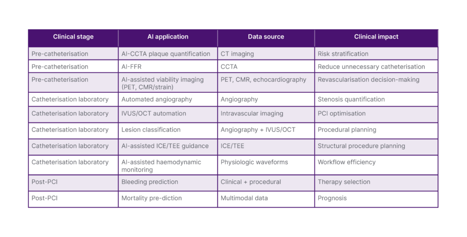
Table 1: Major AI applications in interventional cardiology.
CCTA: coronary CT angiography: CMR: cardiac MRI; FFR: fractional flow reserve: ICE: intracardiac echocardiography; IVUS: intravascular ultrasound; OCT: optical coherence tomography; PCI: percutaneous coronary intervention; TEE: transoesophageal echocardiography.
AI INSIDE THE CATH LAB
While early applications of AI in cardiology focused primarily on diagnostic imaging and risk prediction, AI technologies are increasingly extending into the catheterisation laboratory. Interventional cardiology generates large volumes of structured visual and physiological data during procedures, including angiographic images, intravascular imaging datasets, haemodynamic waveforms, and procedural metadata. These data streams provide an ideal environment for machine learning and deep learning systems capable of assisting clinicians during coronary interventions. Emerging applications include automated angiographic interpretation, intravascular imaging analysis, lesion characterisation, and procedural optimisation. Collectively, these technologies aim to improve procedural precision and reduce variability in complex PCI.16,17
One of the most immediate applications of AI in the catheterisation laboratory is automated interpretation of coronary angiography. Conventional angiographic assessment relies heavily on visual estimation of stenosis severity, which is subject to substantial interobserver variability. Deep learning algorithms trained on large angiographic datasets can automatically identify coronary segments, quantify luminal narrowing, and estimate percent stenosis with reproducibility comparable to expert operators.18 Beyond simple stenosis quantification, AI-assisted quantitative coronary angiography (QCA) platforms have demonstrated improvements in workflow efficiency and consistency. For example, Kwon et al.37 showed that AI-assisted QCA achieved diagnostic accuracy comparable to expert readers while significantly reducing analysis time (1.5±0.9 seconds versus 88.1±35.5 seconds for manual QCA; p<0.001), largely by automating frame selection and minimising operator input.37 Advanced models are also capable of reconstructing 3D coronary anatomy from multiple angiographic projections, allowing more accurate lesion assessment and procedural planning. In addition, AI-based systems can identify complex lesion morphologies, such as bifurcation disease, diffuse atherosclerosis, and chronic total occlusions, potentially providing real-time decision support during PCI.16,37
AI is also transforming the analysis of intravascular imaging modalities, particularly IVUS and OCT. These technologies generate high-resolution cross-sectional images that provide detailed information about plaque composition, vessel dimensions, and stent deployment. However, interpretation requires specialised expertise and time-consuming manual analysis. Deep learning-based segmentation algorithms can automatically delineate vessel borders, quantify plaque burden, and measure key parameters such as minimal lumen area and stent expansion.4 Automated IVUS and OCT analysis platforms, including AI-enabled systems, have demonstrated strong agreement with expert readers while substantially reducing analysis time.
While encouraging, these performance metrics must be regraded with the context that the algorithms underlying automated IVUS and OCT analysis are largely trained on expert-annotated, single-centre datasets whose generalisability may be reduced in clinical settings.38 The accuracy of these measurements largely remains operator and projection dependent, and therefore automation of such tools offers further decision support. Similarly, coronary physiology assessment, whether performed manually or via AI-assisted estimation, is also subject to variability due to hyperaemic agents or measurement technique. However, when these emerging tools are used in conjunction with clinician judgement, they offer avenues for enhancing the patient experience. By enabling rapid and standardised interpretation, AI-assisted intravascular imaging may facilitate broader adoption of image-guided PCI, which has been associated with improved procedural outcomes.39
Automated lesion classification represents another emerging application of AI within the catheterisation laboratory. Coronary lesions vary widely in morphology and complexity, factors that directly influence procedural strategy and outcomes. Machine learning models trained on angiographic and intravascular imaging datasets can classify lesions according to established frameworks such as the American College of Cardiology (ACC)/American Heart Association (AHA) lesion classification, or more advanced phenotypic categories derived from imaging features.40,41 Such AI-based characterisation may help operators anticipate procedural challenges, guide device selection, and determine whether adjunctive techniques, including atherectomy or specialised guidewires, are required.
AI is also being explored to assist with stent sizing and procedural optimisation, two key determinants of PCI success. Algorithms integrating angiographic measurements, intravascular imaging data, and vessel geometry can help estimate optimal stent dimensions and support procedural planning. In addition, automated OCT or IVUS analysis can identify underexpansion, edge dissections, or stent malposition during or after deployment, allowing immediate corrective intervention.39 Beyond these decision-support applications, AI is also contributing to the development of robotic and image-guided PCI systems. While contemporary robotic PCI platforms remain operator-controlled, the integration of AI-driven guidance for tasks such as guidewire navigation, lesion targeting, and device positioning remains investigational. Achieving this in reality requires overcoming well-defined technical barriers, such as compatibility with a broader range of guidewires and over-the-wire, atherectomy, intravascular imaging devices, and active-guide catheter control for tortuous and ostial anatomy.42,43 These emerging technologies suggest a future catheterisation laboratory environment in which computational guidance augments operator performance and procedural safety.40
In addition to coronary interventions, structural heart interventions, including transcatheter aortic, mitral, and tricuspid valve procedures, represent a growing share of procedural volume in many catheterisation laboratories. AI-based software enables real-time registration of fluoroscopy with transoesophageal and intracardiac echocardiography to assist with annular sizing, device selection, and deployment in a manner that is analogous to AI-assisted IVUS/OCT in the coronary realm.7 Both structural and coronary interventions rely heavily on haemodynamic monitoring, and neural network classification has shown to identify real-time damping of aortic pressure waveforms, which can be indicative of serious complications and alters coronary physiology measurements.44 Automated polygraph analysis seeks to benefit the growing popularity of valvular interventions, as well as other structural heart procedures.Together, these tools represent early steps towards an AI-assisted procedural environment in which imaging interpretation, physiological assessment, and device optimisation are increasingly supported by real-time computational analysis. Overall, the integration of AI within the catheterisation laboratory represents a shift towards data-augmented procedural cardiology. Rather than replacing operator expertise, these systems aim to enhance situational awareness, reduce subjectivity in image interpretation, and support more consistent procedural decision-making. As these technologies continue to evolve, the catheterisation laboratory may increasingly incorporate real-time imaging analytics, physiological integration, and procedural guidance algorithms that function alongside clinicians to optimise coronary interventions.
DISCUSSION
Risk Stratification and Outcome Prediction
Beyond imaging interpretation and procedural guidance, AI is increasingly applied to risk stratification and outcome prediction in interventional cardiology. PCI carries risks including bleeding, contrast-induced nephropathy, and mortality, traditionally estimated using risk scores such as Can Rapid risk stratification of Unstable angina patients Suppress ADverse outcomes with Early implementation of the ACC/AHA guidelines (CRUSADE) and Mehran. While clinically useful, these models are limited by fixed variables and linear assumptions, restricting their ability to capture complex patient-level interactions.41,45,46
Machine learning approaches overcome these limitations by integrating large, multidimensional datasets and modelling non-linear relationships. In the context of bleeding risk, AI-based models trained on large clinical registries have demonstrated improved predictive performance compared with traditional risk scores. Machine learning-based models have consistently demonstrated improved discriminative performance compared with traditional risk scores such as CRUSADE and Mehran, with reported C-statistics typically in the range of approximately 0.75–0.85 versus 0.65–0.75 for conventional models. In a comparative effectiveness study that modelled more than three million PCI procedures from the National Cardiovascular Data Registry (NCDR), a machine learning approach improved the prediction of post-PCI major bleeding risk to a C-statistic of 0.82, compared with 0.78 for the existing NCDR model.47 Additionally, a systematic review and meta-analysis also found that machine learning models modestly outperformed traditional risk scores in the discrimination of major adverse cardiovascular outcomes and bleeding risk post-PCI, as demonstrated by consistently higher C-statistics.48 These models incorporate a wide range of variables, including demographic characteristics, comorbidities, laboratory values, procedural details, and pharmacotherapy, enabling more precise individualised risk estimation.41,49
Similarly, AI has shown strong potential in predicting contrast-induced nephropathy, a major complication following coronary angiography and PCI, especially in vulnerable populations such as patients with advanced chronic kidney disease.50 Machine learning models integrating baseline renal function, contrast volume, haemodynamic parameters, and medication exposure have demonstrated superior discrimination compared with conventional scoring systems. Recent studies report significant improvements in predictive accuracy, with machine learning models achieving higher C-statistics than the Mehran score, enabling earlier identification of high-risk patients and targeted preventive strategies such as contrast minimisation and optimised hydration protocols.45,51,52
Beyond complication-specific prediction, AI is increasingly used for global risk estimation and long-term outcome prediction. Models integrating multimodal data, including imaging, physiological measurements, laboratory values, and electronic health records, have demonstrated improved performance in predicting mortality and major adverse cardiovascular events compared with traditional regression-based approaches. For example, machine learning-based models derived from large cohorts such as the Multi-Ethnic Study of Atherosclerosis (MESA) have shown enhanced risk discrimination and reclassification, highlighting the potential of AI to support precision cardiovascular medicine.53
Importantly, AI-based predictive models are not limited to static risk assessment but can enable dynamic, continuously updated risk estimation as new clinical data become available. In addition to guiding individual patient management, these models may support health system-level decision-making by predicting resource utilisation, hospital readmission, and procedural costs. Together, these developments position AI-driven risk stratification as a key component of personalised interventional cardiology.
Regulatory and Implementation Science
As AI technologies transition from research environments into routine clinical practice, regulatory oversight and implementation science have become essential for their safe and effective deployment. Unlike conventional medical devices, AI-based systems, particularly those relying on machine learning, are highly data dependent and may evolve as algorithms are retrained using new datasets, creating unique regulatory and translational challenges.54
In the USA, AI-enabled clinical software is regulated by the FDA under the Software as a Medical Device (SaMD) framework. Several AI platforms used in cardiovascular imaging, including automated coronary CT plaque quantification and AI-derived FFR, have already received regulatory clearance. Most approvals occur through the 510(k) pathway, which requires demonstration of substantial equivalence to existing technologies, while novel systems may be evaluated through the de novo pathway. In Europe, AI medical technologies are regulated under the EU Medical Device Regulation (MDR), which introduced stricter requirements for clinical evidence, risk classification, and post-market surveillance.55,56 Notably, regulatory clearance under both frameworks establishes safety and performance thresholds for market approval, but does not resolve the question of who bears responsibility when an approved software contributes to an adverse outcome and raises major liability issues.
Beyond regulatory approval, robust validation is essential to ensure reliable performance across diverse clinical environments. Contemporary evaluation strategies emphasise multicentre datasets, external validation cohorts, and prospective clinical testing. In addition, concerns regarding algorithm explainability, dataset bias, and performance degradation over time highlight the importance of continuous monitoring after clinical deployment.57-62
Ethical and Practical Challenges
Despite rapid technological progress, the integration of AI into interventional cardiology raises important ethical and practical challenges that must be addressed to ensure responsible clinical adoption. AI systems rely on large datasets, complex computational models, and continuous performance monitoring, creating concerns related to data governance, model stability, generalisability, and clinician preparedness. Addressing these issues is essential for maintaining patient trust, ensuring equitable healthcare delivery, and minimising unintended consequences as AI tools become increasingly integrated into procedural decision-making. Key ethical and implementation challenges associated with AI adoption in interventional cardiology are summarised in Table 2.
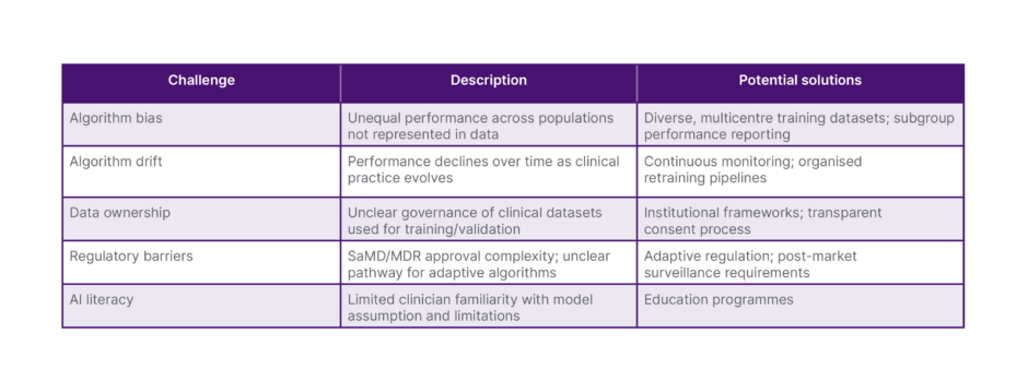
Table 2: Key challenges in clinical AI implementation.
MDR: Medical Device Regulation; SaMD: Software as a Medical Device.
One major challenge involves data ownership and governance. AI systems require large volumes of clinical data for training, validation, and continuous improvement. These datasets often originate from hospitals, imaging archives, and electronic health records containing sensitive patient information. Questions, therefore, arise regarding who owns these data and how they may be used. Healthcare institutions, technology developers, and patients may all have competing claims over data generated during clinical care. In addition, cross-institutional data sharing, essential for developing robust AI models, must comply with privacy regulations and data protection standards. Transparent governance frameworks that define data access, consent processes, and responsibilities for data stewardship will therefore be essential as AI-enabled healthcare ecosystems expand.62,63
Another important concern is algorithm drift, also referred to as model degradation over time. AI models are typically trained on historical datasets reflecting specific clinical environments, imaging protocols, and patient populations. As clinical practice evolves through changes in imaging technology, treatment guidelines, or population demographics, the underlying data distribution may shift. Without periodic recalibration or retraining, this phenomenon can lead to gradual declines in algorithm performance. Continuous monitoring systems, post-deployment auditing, and structured retraining pipelines are therefore necessary to maintain reliable algorithm performance in real-world clinical settings.57
Closely related to algorithm drift is the challenge of generalisability. AI models that demonstrate strong performance in controlled research environments may perform less reliably when applied across different hospitals, geographic regions, or patient populations. Variations in imaging hardware, acquisition protocols, and operator techniques can influence model performance, particularly for imaging-based algorithms used in CCTA, angiography, IVUS, or OCT. Ensuring robust external validation across diverse populations and clinical settings will therefore be critical for the safe and equitable implementation of AI technologies in interventional cardiology.
Lastly, a prominent challenge central to the real-word adoption of AI in interventional cardiology is the question of liability and accountability when AI-assisted decisions lead to patient harm. Responsibility in such cases may be divided among several parties, dependent on the primary cause of patient harm. The device manufacturer may be held accountable if there is a training data flaw or a software defect that caused the algorithm to perform improperly within its intended use. The healthcare institution may be held accountable if the tool was implemented, maintained, or monitored inadequately, or used outside of its validated population or intended use. Ultimately, the treating physician retains the responsibility for the clinical decision regardless of algorithmic input, which makes the upstream parties and AI literacy extremely important. The current regulatory frameworks, SaMD and MDR, establish requirements for pre-market clearance and post-market surveillance, but do not resolve the allocation of liability, especially for adaptive or continuously learning algorithms. While the European legislature primarily pushes accountability onto the provider/manufacturer, the liability issues in the USA are settled through malpractice/tort law due to unclear guidelines. In both instances, the most defensible clinical stance is that AI outputs should be used to guide decision support rather than perform autonomous duties. Clinicians should treat AI-derived measurements and recommendations as inputs to be weighed alongside clinical judgement and independent verification when applicable. As these technologies evolve, the liability frameworks are bound to mature as well and, until then, clear documentation of how AI-derived data is used for clinical decision-making, along with explicit disclosure to patients, may help mitigate the ambiguity of current regulatory frameworks.
Future Directions
As AI technologies mature, the future of interventional cardiology may involve deeper integration of computational intelligence into procedural workflows. Advances in multimodal data integration, digital twin modelling, and real-time decision support may enable the development of AI-assisted catheterisation laboratories capable of continuously integrating imaging, physiological, and clinical data (Supplementary Figure 1).
One emerging concept is the development of real-time AI-assisted catheterisation lab systems. These platforms would continuously analyse live imaging, haemodynamic signals, and procedural data during coronary interventions to provide immediate feedback to operators. For example, AI algorithms could automatically identify coronary anatomy from angiographic frames, detect target lesions, estimate stenosis severity, and suggest optimal imaging projections. When intravascular imaging is used, automated analysis of IVUS or OCT datasets could rapidly quantify minimal lumen area, plaque burden, and stent expansion in near real time. By functioning as an intelligent procedural assistant, such systems may enhance situational awareness, reduce operator cognitive load, and improve procedural consistency.4,37
Another promising area involves the development of cardiovascular digital twins, computational replicas of individual patients that integrate anatomical, physiological, and clinical data. These models could combine coronary imaging datasets with haemodynamic parameters and patient-specific clinical variables to simulate disease progression and treatment responses. In interventional cardiology, digital twins may allow clinicians to test alternative revascularisation strategies before performing a procedure, for example, by predicting the physiological impact of stenting a particular lesion or estimating post-procedural FFR. Although still largely experimental, digital twin frameworks represent a potential step towards predictive and personalised coronary intervention.64
A key enabling technology for these future systems is multimodal data fusion. Modern cardiovascular care generates diverse information streams, including CCTA, invasive angiography, intravascular imaging, physiological measurements such as FFR or instantaneous wave-free ratio, and extensive clinical data from electronic health records. AI models capable of integrating these heterogeneous datasets may provide more comprehensive insights than models relying on single modalities, supporting procedural planning, risk prediction, and long-term management decisions.4,8
Finally, AI may further expand the capabilities of robotic-assisted PCI. Current robotic platforms allow operators to manipulate devices remotely, reducing radiation exposure and improving operator ergonomics. Future systems may incorporate AI-driven guidance algorithms to assist with tasks such as guidewire navigation, lesion targeting, and device positioning. Although fully autonomous PCI remains a theoretical possibility, partial automation and AI-assisted device navigation may enhance procedural precision and safety.42,43
Taken together, these emerging developments suggest that AI in interventional cardiology may evolve from isolated analytical tools to integrated procedural ecosystems. Real-time decision support, patient-specific modelling, multimodal data integration, and advanced robotics may progressively transform how coronary interventions are planned and performed.
CONCLUSION
AI is rapidly reshaping interventional cardiology, evolving from tools primarily used for imaging analysis into broader platforms for procedural intelligence and clinical decision support. Advances in automated imaging interpretation, physiological modelling, and predictive analytics are enabling more precise patient selection, improved procedural planning, and enhanced outcome prediction across the continuum of coronary artery disease management.
Importantly, the role of AI is progressively shifting from a non-invasive imaging gatekeeper that refines patient selection to a procedural co-pilot capable of assisting clinicians during coronary interventions. Rather than replacing physician expertise, these technologies are designed to augment human judgement by improving image interpretation, reducing procedural variability, and supporting data-informed decision-making.
As computational capabilities continue to advance, the catheterisation laboratory may increasingly evolve into a data-driven environment in which imaging, physiology, and clinical information are integrated in real time to support more personalised and optimised coronary interventions.