Key Summary:
- Visual assessment alone may miss early pressure injuries in dark skin.
- Four technology supported approaches detected changes before visible signs.
- Evidence quality and multimodal assessment remain important research gaps.

PRESSURE injury detection in dark skin may improve when clinicians supplement visual checks with objective technologies.
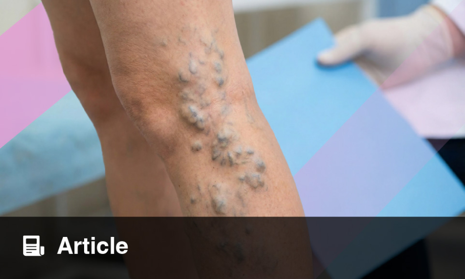
Early pressure injuries may be harder to recognize in people with darker skin tones because erythema, blanching, and pink or purple discoloration can be less visible. Delayed identification may allow reversible Stage 1 damage to progress to deeper tissue injury, infection, longer hospitalization, and greater morbidity.
Researchers mapped strategies used to identify pressure injuries in adults with dark skin tones in acute care. They searched five databases for English language primary research published since 2017. Of 1,353 records identified, 10 studies met the eligibility criteria. All originated in the U.S. and included participants with dark skin tones, although definitions of skin tone varied.
The studies covered four approaches: thermographic imaging, subepidermal moisture sensors, image processing and colorimetry, and enhanced assessment with specialized lighting. Across these categories, objective tools detected tissue changes before visible signs appeared and generally maintained performance across diverse pigmentation levels.
Thermography identified temperature abnormalities before pressure injuries became visually apparent. Subepidermal moisture measurements detected tissue changes days earlier than visual examination in some studies. One study reported detection 3 to 10 days before visible signs, while another found that visual confirmation took an average of 4.3 days.
In a quality improvement study involving 140 critical care patients, hospital acquired pressure injuries fell from 8.9% before subepidermal moisture scanning to 0% during implementation. Image processing and colorimetry provided more reproducible measurements of erythema and wound boundaries. Enhanced lighting also revealed changes not seen under standard white light, although benefits varied in darker skin tones.
For clinicians, the findings support considering temperature, moisture, pain, and tissue firmness or softness alongside color change during skin assessment. Objective tools may strengthen pressure injury detection in skin of color, particularly in acute and critical care settings where immobility and physiologic instability increase vulnerability.
However, the review did not formally appraise study quality, and no study evaluated a multimodal strategy. Routine visual and tactile assessment was also underrepresented. Costs and infrastructure may limit adoption, while artificial intelligence studies did not describe safeguards against skin tone related algorithmic bias. The authors called for stronger studies of both scalable bedside methods and technology supported assessment before widespread integration.
Reference
Mbuzi VB et al. Skin assessment strategies for identification of pressure injuries in dark-skin-tone patients: A scoping review. Healthcare (Basel). 2026;14(16):2655.
Featured Image: Seventyfour on Adobe Stock.
Each article is made available under the terms of the Creative Commons Attribution-Non Commercial 4.0 License.