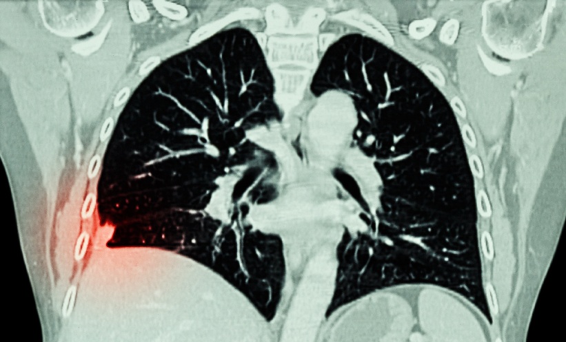
LUNG CANCER screening delivered through a cloud-native imaging network enabled more than one million low-dose CT (LDCT) scans to be interpreted since 2020, while maintaining rapid reporting at national scale, according to a new report.
The authors suggested the infrastructure could provide a framework for other imaging-based screening programmes if adapted to their specific clinical requirements.
Lung cancer, the leading cause of cancer death worldwide, can be detected earlier through LDCT screening, which has been shown to reduce lung cancer mortality. The NHS England Lung Cancer Screening Programme was described as the largest national LDCT implementation to date, having issued more than 2.5 million invitations, diagnosed 7,193 lung cancers and detected 63.1% of cases at stage I during its first five-year evaluation.
Three-Part Model Enabled National Lung Cancer Screening
The report identified three components as central to delivering the programme at scale. First, a cloud-native, vendor-agnostic imaging information technology platform addressed fragmentation across NHS picture archiving and communication systems, enabling distributed image acquisition, centralised expert interpretation and standardised structured reporting.
Second, a virtual national specialist network of 180 consultant thoracic radiologists worked within a single reporting environment rather than through a transactional teleradiology contract. According to the authors, this model supported subspecialist reporting, embedded peer review, discrepancy logging and named consultant accountability.
Third, vendor-agnostic integration of artificial intelligence for lung nodule detection and volumetry enabled network-level monitoring for algorithm drift and population bias after deployment.
Together, these three components supported interpretation of more than one million LDCT studies since 2020 and returned more than 99% of 41,000+ monthly LDCT scans within 72 hours.
Why Imaging-Based Screening Required a Different Model
The authors argued that imaging-based screening differs from biochemical and cytological screening because image interpretation requires subspecialist expertise, imaging data are large and distributed across multiple systems, and decisions are made over repeated screening rounds spanning many years.
They suggested these characteristics made both generalist in-house reporting and traditional teleradiology poorly suited to population screening, requiring a different organisational approach.
Lessons Could Extend Beyond Lung Cancer Screening
The report suggested the underlying model could be adapted for other imaging-based screening programmes, including breast screening, prostate MRI, CT colonography and, potentially, cardiac CT. However, the authors emphasised that clinical pathways, evidence bases and reporting requirements differ between programmes, with some applications remaining investigational.
They concluded that imaging-based screening should be commissioned as integrated clinical infrastructure rather than through separate technology and workforce procurement. The authors suggested the architecture developed for the NHS lung cancer screening programme could provide a promising, programme-specific foundation for future imaging-based screening initiatives.
Reference
Hare S et al. Scaling AI-enabled imaging-based screening: lessons from reporting for the NHS England Lung Cancer Screening Programme. Clin Radiol. 2026;DOI:10.1016/j.crad.2026.107429.
Featured image: Stockdevil on Adobe Stock