Abstract
The incidence of mechanical complications following acute myocardial infarction has decreased significantly with contemporary early reperfusion strategies. Nevertheless, when present, they remain life-threatening conditions requiring urgent intervention. Although surgical repair continues to be the standard of care for ventricular septal rupture in haemodynamically stable patients, emerging evidence supports transcatheter percutaneous closure as a viable alternative in critically ill, haemodynamically unstable patients. Herein, the authors report the case of a 65-year-old female with a prior medical history of arterial hypertension and Type 2 diabetes who presented with subacute anterior ST-segment elevation myocardial infarction. The patient had not sought medical attention at the time of the initial event and was admitted to the institution several days after symptom onset with clinical features of post-infarction heart failure. A transthoracic echocardiogram revealed a ventricular septal rupture, confirming the presence of a potentially lethal mechanical complication. Following multidisciplinary heart team evaluation, transcatheter percutaneous closure was deemed the most appropriate therapeutic strategy given the patient’s haemodynamic instability, heart failure status, and dependence on vasopressor and ventilatory support, which precluded surgical repair. The procedure was performed successfully, and at the time of this report, the patient remains cardiovascularly asymptomatic with no hospital readmissions, demonstrating that percutaneous closure is a feasible and safe intervention in this clinical setting.
Key Points
1. Recurrent malignant pericardial effusion is a life-threatening complication in patients with advanced cancer, with high recurrence rates following pericardiocentesis alone.2. Simplifying the procedure as much as possible and limiting the number of implanted stents by using a stepwise provisional strategy remains the recommended strategy for the majority of bifurcation PCI.
3. In this review article, the authors propose the Patient, Lesion, Operator, Technique, and Outcomes (PLOTO) framework to approach bifurcation PCI, which focuses on the five titular components. This model aims to tailor interventions to individual patient needs and help select the most appropriate technique to optimise outcomes.
CASE PRESENTATION
A 65-year-old female patient, originating from a rural area with limited access to healthcare, presented to the authors’ institution following transfer from a local medical facility. She reported a 15-day history of chest pain of cardiovascular aetiology, for which she had not previously sought medical attention. She was transferred with a working diagnosis of post-infarction heart failure and was admitted to the ICU with a presentation consistent with acute decompensated heart failure. Her past medical history was notable for arterial hypertension and Type 2 diabetes. Physical examination revealed a pansystolic cardiac murmur, and a 12-lead ECG demonstrated pathological Q waves in leads V1 through V4 with persistent ST-segment elevation, consistent with a subacute anterior wall myocardial infarction. Transthoracic echocardiography revealed an apical septal ventricular wall rupture with a resultant ventricular septal defect and akinesia of the entire anterior wall, consistent with a subacute anterior myocardial infarction. Subsequent coronary angiography demonstrated complete occlusion of the mid left anterior descending (LAD) artery, along with severe stenosis of the proximal and mid segments of the right coronary artery.
Despite the institution of escalating vasoactive and inotropic support, comprising noradrenaline, vasopressin, and levosimendan, the patient demonstrated no meaningful clinical improvement, with progressive haemodynamic deterioration. Formal calculation of the pulmonary-to-systemic flow ratio was not performed, as the patient’s clinical status left little diagnostic ambiguity; the haemodynamic compromise was clearly attributable to the underlying mechanical complication. Given the severity of the patient’s progressive haemodynamic instability, the multidisciplinary Heart Team convened and reached a consensus that emergency transcatheter closure of the ventricular septal defect represented the most appropriate and expedient therapeutic strategy.
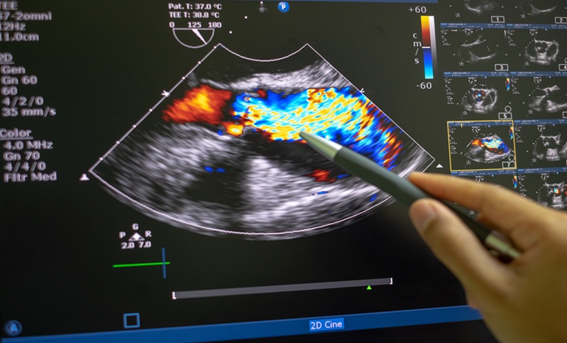
Haemodynamic support was initially established with an intra-aortic balloon pump. An arteriovenous circuit was created from the right internal jugular vein to the right femoral artery, traversing the ventricular septal defect, using a 0.035” × 260 cm hydrophilic guidewire. Device selection was determined by institutional availability at the time of the procedure. Given the post-infarction aetiology of the ventricular septal defect, the surrounding myocardial tissue was expected to be friable, necrotic, and poorly defined at its margins, characteristics inherent to this clinical entity that preclude precise anatomical delineation and mandate a more generous device oversizing strategy compared to congenital defects. The defect was measured at 9 mm by both transthoracic echocardiography and fluoroscopic angiography, with concordant findings across both modalities. In accordance with current practice for post-infarction ventricular septal rupture, wherein an oversizing margin of 2–4 mm above the maximum measured diameter is recommended to account for tissue friability and the potential for defect enlargement, a #26 Hyperion™ VSD occluder (Comed BV, Heerenveen, the Netherlands) was selected. This device comprises a self-expandable, double-disc nitinol mesh framework with integrated polyester fabric, designed to promote thrombogenesis and accelerate neo-endothelialisation at the implant site. Through a 12-Fr delivery sheath, the #26 Hyperion occluder device was advanced and successfully deployed across the defect (Figure 1). Prior to device release, stable and secure positioning was confirmed by performing the Minnesota manoeuvre, a standardised assessment technique consisting of the application of gentle, controlled push-pull traction on the delivery cable whilst simultaneously evaluating device position under fluoroscopic and echocardiographic guidance. Absence of device displacement, maintained disc apposition against both the left and right ventricular septal surfaces, and absence of interference with adjacent valvular structures were confirmed before final device release was performed.
Figure 1: Emergency transcatheter VSR closure with Hyperion™ occluder.
A) Transthoracic echocardiographic colour Doppler imaging demonstrating a post-STEMI ventricular septal rupture with a left-to-right intracardiac shunt. B) Echocardiographic assessment revealing extensive anterior wall akinesia with apical aneurysm formation. C) Coronary angiography demonstrating atherothrombotic occlusion of the mid left anterior descending artery. D) Coronary angiography demonstrating severe atherosclerotic disease involving the proximal and mid segments of the right coronary artery. E–F) Left ventriculography confirming the ventricular septal rupture with evidence of a left-to-right shunt. G) Establishment of an arteriovenous circuit across the ventricular septal defect using a 0.035″ hydrophilic guidewire. H) Advancement and positioning of the Hyperion™ #26 occluder device across the ventricular septal defect. I) Successful closure of the ventricular septal rupture confirmed through Minnesota manoeuvre and post-deployment left ventriculography demonstrating no residual shunting. J) Fluoroscopic imaging showing the asymmetric Hyperion #26 occluder device deployed across the ventricular septal rupture. K) Percutaneous coronary intervention of the right coronary artery with successful deployment of a 3.5 × 48 mm drug-eluting stent. L) Follow-up transthoracic echocardiography demonstrating appropriate device positioning with no evidence of residual left-to-right shunting.
Hyperion™ #26 occluder: Comed BV, Heerenveen, the Netherlands.
STEMI: ST-segment elevation myocardial infarction; VSR: ventricular septal rupture.
Concomitant percutaneous coronary intervention of the right coronary artery was performed, with successful deployment of a 3.5 × 48 mm drug-eluting stent, achieving Thrombolysis in Myocardial Infarction (TIMI) Grade 3 flow post-procedure. The decision was made not to intervene on the LAD artery, given the echocardiographic evidence of extensive akinesia with marked wall thinning and aneurysmal remodelling of the subtended myocardial territory, in keeping with established, irreversible myocardial injury secondary to a subacute total occlusion. In the acute setting, the clinical context did not warrant formal myocardial viability assessment of the LAD territory, as the morphological findings were consistent with completed infarction with no evidence of salvageable myocardium. Furthermore, the elevated risk of reperfusion-related myocardial injury in the setting of subacute occlusion with established aneurysmal changes further supported a conservative approach.
At follow-up, given the patient’s sustained clinical stability, a goal-directed pharmacological management strategy was maintained, and intervention on the LAD territory is not currently indicated.
At 1 year of clinical follow-up, the patient remains in a satisfactory cardiovascular status, reporting no symptoms attributable to either the procedure or her underlying condition. Serial echocardiographic assessment has demonstrated no evidence of residual shunting, and she has required no further reintervention to date.
Her clinical course throughout thisperiod has been free of procedure-related or disease-related complications, reflecting a favourable long-term outcome following transcatheter ventricular septal defect closure.
DISCUSSION
Contemporary early revascularisation strategies have significantly reduced the incidence of ventricular septal rupture (VSR) following acute myocardial infarction. Nevertheless, when present, VSR remains a life-threatening mechanical complication. Natural history data consistently demonstrate a mortality rate of approximately 24% within the first 24 hours of presentation, rising to 46% at 1 week and between 67–82% at 2 months in the absence of mechanical intervention. Lemery et al.1 further reported a 30-day survival rate of only 24% among medically treated patients, compared with 47% among those managed surgically,2 underscoring the critical importance of prompt mechanical closure.
Without definitive intervention, mortality approaches 87–100% within 2 months of diagnosis, rendering VSR one of the most lethal complications of acute myocardial infarction.3 Delayed surgical repair, when feasible, continues to be regarded as the standard of care for post-ST-segment elevation myocardial infarction VSR. However, percutaneous transcatheter closure represents a viable therapeutic alternative in patients deemed to have prohibitive surgical risk or refractory haemodynamic instability, as illustrated in the case presented herein.4 Although in-hospital mortality following transcatheter closure of post-infarction ventricular septal rupture remains substantial, with reported rates of up to 58%,5 outcomes are demonstrably more favourable when intervention is performed in the subacute setting, where 30-day survival rates of up to 81% have been reported following percutaneous ventricular septal rupture closure.3,4
CONCLUSION
Post-STEMI ventricular septal rupture remains a life-threatening mechanical complication associated with exceedingly high mortality rates if left untreated. Transcatheter percutaneous closure should be strongly considered as a viable therapeutic alternative whenever surgical repair is deemed unfeasible or carries prohibitive risk.