Interview Summary
Epilepsy is a chronic neurological disorder characterised by recurrent, unprovoked seizures that can significantly affect morbidity, mortality, and quality of life (QoL). Around 30% of people with epilepsy develop drug-resistant epilepsy (DRE), meaning seizures persist despite treatment with two or more adequate trials of antiseizure medications (ASM). For these individuals, alternative therapeutic approaches are often required. Vagus nerve stimulation (VNS) is a well-established adjunctive neuromodulation therapy that has been used for more than 3 decades. Clinical evidence shows that it can reduce seizure frequency and burden, extend seizure-free intervals, and may also contribute to lowering the risk of sudden unexpected death in epilepsy (SUDEP).Evidence suggests that VNS works through the vagal afferent network to influence key brainstem nuclei and downstream brain circuits involved in seizure generation and propagation. Stimulation of the nucleus tractus solitarius (NTS) and locus coeruleus (LC) appears to increase the release of monoamines such as noradrenaline (NA) and 5-hydroxytryptamine (5-HT), better known as serotonin, which in turn modulate neurotransmission.
Here, Raman Sankar, Professor of Neurology and Paediatrics and Chief of Paediatric Neurology, David Geffen School of Medicine, University of California, Los Angeles, USA, proposes and outlines the evidence for a monoamine hypothesis of VNS function. He also argues that a clearer understanding of this mechanism of action (MOA) can help clinicians work with patients to make more informed treatment decisions.
INTRODUCTION
Epilepsy is a chronic neurological condition characterised by a predisposition to unprovoked seizures.1 It can have a significant impact on morbidity, mortality, and QoL.1-3 Around 30% of people living with the condition have DRE, meaning they fail to achieve freedom from seizures after trying two or more ASMs at an efficacious dose.3,4
VNS is an adjunctive neuromodulatory therapy that has been shown to be efficacious in DRE.5 It is a long-established non-pharmacological intervention that involves a small pulse generator being surgically implanted under the skin of the chest. A generator transmits electrical signals to the vagus nerve via a lead wire.6 The therapy has been shown to reduce seizure frequency and severity, improve QoL and recovery, and, potentially, reduce the risk of SUDEP.6-8
Our understanding of the VNS MOA continues to evolve, and Sankar believes it is important to “put what we do know up front.” This enables “more scientifically grounded conversations” about treatment selection with clinicians and patients alike. “Doctors who prescribe ought to be well informed on what is available,” he added.
THE VAGAL AFFERENT NETWORK AND EPILEPSY
The literature suggests VNS is a network therapy that, in responders, modulates brain synchrony towards a less epileptogenic state via the vagal afferent network.9 Sankar explained that the vagal afferents primarily project to the NTS, which sends fibres, both directly and via the nucleus paragigantocellularis and nucleus prepositus hypoglossi, to other brainstem nuclei that modulate subcortical and cortical activity (Figure 1).10,11 These include the LC, the brain’s primary source of NA, and the dorsal raphe nuclei (DRN), the main producer of serotonin.10-13
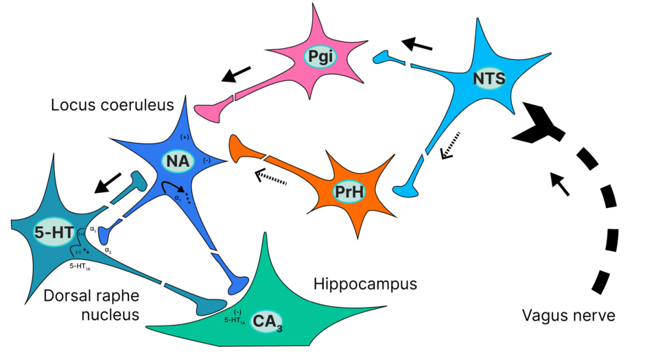
Figure 1: Connectivity of the vagus nerve to critical brainstem nuclei.10
5-HT: serotonin; CA3: cornu ammonis 3; NA: noradrenaline; NTS: nucleus tractus solitarius; Pgi: nucleus paragigantocellularis; PrH: nucleus prepositus hypoglossi.
The thalamus also plays a key role in facilitating seizure propagation, likely due to its extensive reciprocal cortical and limbic connectivity as well as its demonstrated role in promoting cortical synchronisation during cognition.14 The cortex is most often the brain region current diagnostic approaches localise focal seizures to. However, the International League Against Epilepsy (ILAE) notes that focal seizures may originate in subcortical structures as well.15
MONOAMINERGIC MODULATION OF LIMBIC AND THALAMOCORTICAL CIRCUITRY
VNS reduces seizures by disrupting the hypersynchronous state typical of seizures.9 EEG data from people who have received and responded to the therapy show neural desynchronisation in both acute and long-term states.9,16,17 Evidence, primarily from animal research, suggests it does this through bottom-up monoaminergic modulation of the limbic and thalamocortical circuitry that propagate seizures, explained Sankar.
VNS stimulates the brainstem nuclei (NTS), with the signal propagating through a connected system that includes the LC, limbic, and thalamocortical networks.10,18,19 There is growing evidence to suggest that VNS enables neurogenesis and plasticity in the hippocampus and alters neuronal excitability within the amygdala and hippocampus.11,20 Studies have also shown it increases the seizure threshold of neurons in the amygdala.21 Others have suggested that VNS improves thalamic metabolism and seizure response,11 while increasing cortical receptor density of gamma-aminobutyric acid Type A receptors.22,23
Sankar explained that, in a 1998 rat study, VNS efficacy was blocked by the lesioning of the LC, suggesting the nuclei are at the start of a cascade effect that results in the limbic and thalamocortical circuitry modulation.24 “Krahl did a very simple thing: he had animals that were implanted with VNS, and they were responding. He then destroyed the LC with a neurotoxin, and VNS stopped working. The next question has to be: what’s going on in the LC?” he said, reiterating the nuclei’s role both in producing NA, and in activating the DRN, which produces 5-HT.
Highlighting the relationship between VNS, the LC, and monoamines such as NA and 5-HT, he pointed to another rat study, in which microdialysis demonstrated increased extracellular NA in the hippocampus and the cortex following VNS therapy.25 Others have found increased hippocampal NA to be a biomarker of VNS efficacy, and that NA depletion or LC damage accelerates the rate of amygdala kindling, a model for mesial temporal lobe epilepsy.26-28 Researchers have also found reduced α1-adrenergic receptors in surgical tissue resected from patients undergoing temporal lobectomy for focal DRE.29 In terms of serotonin, the optogenetic activation of 5-HT neurons in the DRN is anticonvulsant and suppresses respiratory arrest in the DBA/1 mouse SUDEP model.30 Sankar also pointed to a study that found both NA and 5-HT were necessary to elicit VNS-directed cortical plasticity in rats.31
Based on such findings, VNS is believed to act first on the LC, and then indirectly on 5-HT neurons in the DRN, leading to a marked increase in NA and 5-HT transmission. It is this modulation of monoamines, derived from modulation of the vagal afferent network via the NTS, that is thought to impact changes in the limbic and thalamocortical circuitry, and promote the known effects of VNS (Figure 2).9,10,20,22,23,32,33
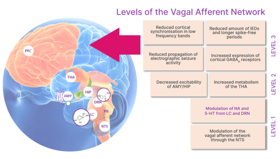
Figure 2: Bottom-up modulation of the vagal afferent network.9,10,20,22,23,32,33
5-HT: serotonin; AMY: amygdala; GABAA: gamma-aminobutyric acid type A; DRN: dorsal raphe nucleus; HIP: hippocampus; IED: interictal epileptiform discharges; LC: locus coeruleus; NA: noradrenaline; NTS: nucleus tractus solitarius; PB: parabrachial nucleus; PFC: prefrontal cortex; THA: thalamus.
Image provided by LivaNova.
WHY MOA MATTERS
A deeper understanding of VNS MOA is important, said Sankar, because it can help to guide complex treatment selection decisions in DRE. Multiple ASMs are available and, in recent years, additional forms of neuromodulation, such as intracranial responsive neurostimulation and deep brain stimulation, have emerged. As VNS provides broad network modulation without intracranial surgery, it may be considered a logical, early intervention immediately following the failure of two to three ASMs, before considering more invasive options. “Everyone is talking about responsive neurostimulation and deep brain stimulation in the anterior nucleus of the thalamus, but memories are short in medical history,” he said, pointing out that while VNS has been available for more than 30 years, it remains an important part of the treatment landscape. “If you are going to select neurostimulation, you are going to find that not every patient wants detailed, invasive monitoring, and not everybody wants electrodes put into their brain permanently. If you do not have to go inside the brain, people are more comfortable with that,” said Sankar.
He went on to say that while seizure reduction tends to be the primary endpoint in clinical trials, it was important for healthcare professionals to take a wider view. “Around the turn of the century, important papers showed that whether you have seizure reduction of 45% or 60%, it is not a meaningful difference,” he said.34 When patients are asked about the major QoL detractors, he went on, many speak about co-morbidities, such as depressive symptoms.34 He pointed to a 2020 review article in which six of the nine included studies reported that VNS had a positive impact on depressive symptoms. In addition, eight of the nine did not find any correlation between seizure reduction and the amelioration of depressive symptoms.35 Simply reducing seizures may not be sufficient to reduce comorbid depression, then, Sankar explained, adding that VNS had a clear mechanistic advantage, through the modulation of serotonin.
With more information about the VNS MOA, healthcare professionals can have clearer, more informative conversations with their patients, Sankar went on. It enables them to explain how different interventions target different pathways, and that VNS may modulate NA and 5-HT systems that innervate the entire network involved in seizure propagation, including regions involved in seizures.10 This can help patients understand why other medications, which target only a single node in the network or a single neurotransmitter, may have failed, and work with their healthcare team to make informed treatment decisions.
A greater understanding of how VNS works can also help guide decisions around treatment targets. It may act upon serotonergic pathways that “may not have been exploited” in some patients living with DRE, Sankar said. Evidence supporting the importance of serotonin in seizure control, he explained, comes from drugs such as fenfluramine. The agent, which increases serotonin release and blocks its reuptake, has demonstrated strong efficacy in some rare forms of epilepsy and has also been associated with SUDEP risk reduction in patients with Dravet syndrome.28-38 Sankar highlighted that the agent has a narrow indication, only being licensed for seizures associated with Dravet and Lennox-Gastaut syndromes. 6,39 As such, VNS provides a way to target the critical serotonergic pathways in a broader population of people living with DRE, he explained.
CONCLUSION
VNS remains an important therapeutic option for people with DRE, offering seizure frequency and severity reductions in responders.6 As with most therapies, the precise mechanism of VNS is challenging to confirm in the human condition. However, growing evidence supports that VNS is a network therapy. It engages the vagal afferent network and influencing key brainstem nuclei that regulate widespread cortical and subcortical circuits involved in seizure generation and propagation.10,11 Activation of NTS and, in turn, the LC, appears to initiate a cascade that increases NA and 5-HT transmission, ultimately modulating limbic and thalamocortical circuitry and promoting neural desynchronisation.10,11 Understanding the MOA of VNS has practical implications for clinical decision-making and supports clearer communication with patients around treatment selection and the avoidance of QoL-reducing comorbidities, such as depression.