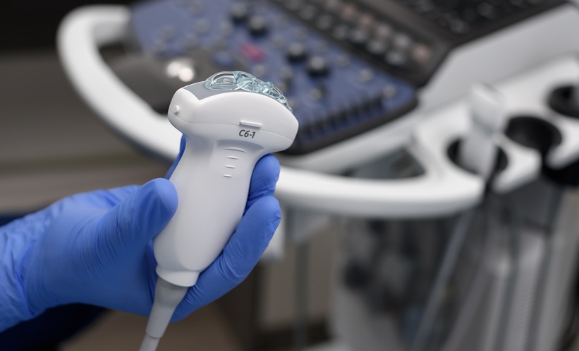
TRANSIENT elastography combined with non-invasive biomarker scores could help detect and stage liver fibrosis in people with thalassaemia, according to a systematic review.
The analysis found that combining transient elastography (TE) with the Fibrosis-4 (FIB-4) score showed promise for identifying early fibrosis, while TE-based approaches also demonstrated potential for detecting cirrhosis.
Finding Alternatives to Liver Biopsy
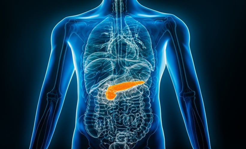
People with thalassaemia are at risk of liver fibrosis as a result of factors including iron accumulation from repeated blood transfusions and increased intestinal iron absorption.
Liver biopsy remains the reference standard for fibrosis assessment, but its invasive nature makes repeated testing impractical.
Researchers therefore systematically reviewed evidence examining radiological imaging and serum biomarker approaches for detecting and staging fibrosis in patients with thalassaemia.
Nine studies involving 500 patients were included, assessing 12 diagnostic approaches. Despite searching across radiological modalities, TE was the only imaging technique evaluated in the eligible studies.
Combining Imaging With Biomarkers
For detecting fibrosis of METAVIR stage F1 or greater, TE combined with FIB-4 achieved an area under the receiver operating characteristic curve of 0.74.
The combination had a sensitivity of 85%, although specificity was considerably lower at 36.5%.
TE alone demonstrated variable performance for early fibrosis, with reported AUC values ranging from 0.34 – 0.73. For advanced fibrosis, results also varied according to the reference standard used.
The researchers noted that some serum biomarkers produced apparently strong diagnostic performance when compared with surrogate reference standards. However, these findings should be interpreted cautiously because the reference tests themselves did not consistently perform well against liver biopsy.
Potential Role in Cirrhosis Detection
TE appeared more promising for detecting cirrhosis. Cut-offs between 11–13 kPa and approaches combining TE with either APRI or FIB-4 demonstrated encouraging diagnostic accuracy for METAVIR F4 disease.
The ability to assess liver fibrosis non-invasively could be particularly valuable in thalassaemia, where patients require long-term monitoring and repeated biopsy is undesirable.
However, the evidence base remains limited. None of the 12 diagnostic tests was evaluated in more than three studies for an individual fibrosis stage, and differences between reference standards prevented researchers from pooling diagnostic performance statistically.
Further imaging-focused research using liver biopsy as a consistent reference standard is needed to determine how TE and other radiological techniques should be incorporated into routine fibrosis surveillance.
Reference
Chandra P et al. Radiologic imaging and biomarker indices in detecting liver fibrosis in thalassemia: a systematic review of diagnostic accuracy. Egypt J Radiol Nucl Med. 2026;57:197. doi:10.1186/s43055-026-01872-3.