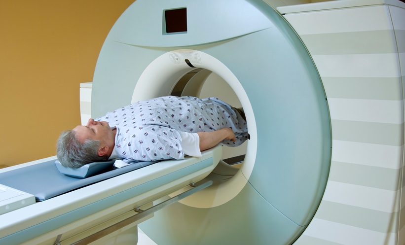
COMBINING multiparametric MRI with clinical information may improve prediction of prostate cancer upgrading after surgery, a retrospective study suggests.
The analysis examined men whose biopsies showed Gleason scores of 3+3 or 3+4, corresponding to Grade Groups 1 and 2. More than half had a higher grade identified when their prostate was examined after radical prostatectomy, highlighting the potential for biopsy findings to underestimate disease severity.
Comparing Biopsy and Surgical Findings
Researchers evaluated clinical and MRI data from 145 patients at a single institution. All underwent prostate biopsy followed by radical prostatectomy, allowing comparison between the grade assigned to biopsy samples and the final surgical specimen.
Of the participants, 83 had a biopsy Gleason score of 3+3 and 62 had a score of 3+4. Overall, 81 patients, or 55.9%, experienced upgrading after surgery.
Among those with a biopsy score of 3+3, 53 patients, or 63.9%, were upgraded to at least 3+4. Among those initially graded 3+4, 28 patients, or 45.2%, were upgraded to at least 4+3.
Upgrading in this setting means that surgical pathology identified a higher grade than biopsy; it does not necessarily indicate that the cancer became more aggressive between the two procedures.
MRI Adds Predictive Information
The researchers compared three approaches: a clinical model, an MRI model, and a combined model incorporating both sets of information.
Clinical variables included age, prostate-specific antigen measures, prostate volume, and biopsy-related information. Imaging variables included tumour diameter and location, the MRI grade of extraprostatic extension, and PI-RADS assessment.
The clinical model achieved an area under the receiver operating characteristic curve of 0.621, compared with 0.707 for the MRI model. Combining clinical and imaging information increased this to 0.722, a statistically significant improvement over clinical assessment alone (p=0.017).
Patients whose cancers were upgraded also had a greater proportion of lesions at the prostate base and higher MRI grades of extraprostatic extension. Nevertheless, the combined model’s predictive performance remained moderate.
Implications for Risk Assessment
The findings suggest that MRI could add useful information when discussing whether biopsy findings adequately reflect a patient’s cancer. Better prediction could potentially inform decisions about surveillance and definitive treatment.
However, this study did not demonstrate that using the model improves treatment outcomes or reduces undertreatment. Its retrospective design, single-centre population, and inclusion only of patients who underwent surgery also limit generalisability, particularly to men choosing active surveillance.
The authors additionally noted differences between imaging and pathological appearances and potential variation in image interpretation. Further validation is needed to establish how reliably the approach performs across clinical settings.
Reference
Zhang J et al. Value of multiparametric MRI in predicting postoperative upgrading of prostate cancer with biopsy Gleason Grade Groups 3+3 and 3+4. BJR Open. 2026. doi:10.1093/bjro/tzag022.