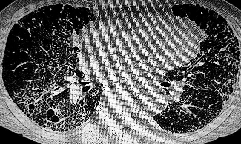
A U-I-P score developed from three high-resolution CT (HRCT) features identified histopathological usual interstitial pneumonia (UIP) features and predicted disease progression in smokers with fibrotic idiopathic interstitial pneumonia (IIP), according to a retrospective multicentre study.
UIP is a pattern of lung scarring linked to progressive fibrosis. Recognising it on CT scans can be challenging in smokers because emphysema and smoking-related fibrosis may obscure typical imaging features of UIP.
The imaging-based U-I-P score was designed to improve identification of UIP pathology in patients whose CT appearances were atypical or when lung biopsy was not available. For healthcare professionals, this could provide additional support when assessing smokers with fibrotic IIP whose imaging findings make diagnosis more difficult. Researchers found the score remained a strong predictor of both histopathological UIP features and subsequent disease progression across independent patient cohorts.
Simple Imaging Score Showed Strong Predictive Performance
The study included two biopsy-proven cohorts of smokers with fibrotic IIP. The derivation cohort comprised 242 patients from a nationwide registry, while the validation cohort included 126 patients from a single-centre database collected between 2008 and 2013.
Investigators assessed three predefined HRCT findings: upper-lobe irregular lines, irregularity of the pleural surface, and pleural-based basal reticulation. Each feature was assigned one point, creating a U-I-P score ranging from 0 to 3.
The cohorts were comparable, with median ages of 65 and 64 years, and men accounting for 88% and 87% of participants, respectively. Histopathological UIP features were identified in 208 of 242 patients (86%) in the derivation cohort and 109 of 126 patients (87%) in the validation cohort.
The U-I-P score independently predicted histopathological UIP features in both cohorts, achieving area under the curve values of 0.83 and 0.89 (both p<0.001). Bootstrap validation demonstrated minimal optimism, supporting the model’s stability.
Score Also Identified Patients at Greater Risk of Progression
In the validation cohort, the U-I-P score independently predicted disease progression at both one and two years (p=0.002 and p=0.001, respectively).
The findings suggested the U-I-P score could serve as a practical imaging biomarker for identifying smokers with UIP-type fibrosis and those with a greater likelihood of disease progression, including patients whose CT findings did not meet typical UIP classification. The investigators indicated the approach may support clinical assessment when biopsy was unavailable, although the study was retrospective and its findings were derived from biopsy-proven cohorts.
Reference
Egashira R et al. A CT-based score for predicting histopathological usual interstitial pneumonia features and predicting disease progression in smokers with fibrotic idiopathic interstitial pneumonia. Eur Radiol. 2026;DOI:10.1007/s00330-026-12741-y.
Featured image: DOUGLAS on Adobe Stock