Abstract
Background: The prevalence of chronic kidney disease (CKD) has been increasing globally (9.1%) and in Kenya (10.6%). There is emerging evidence of an increased risk of cervical dysplasia and cervical cancers in patients with CKD. Cervical cancer screening is part of secondary prevention of cervical cancer that leads to early detection and treatment of premalignant lesions. Despite the high prevalence of CKD, little is known about the patterns and prevalence of cervical cytological abnormalities in this subset of the population in Moi Teaching and Referral Hospital (MTRH), Eldoret, Kenya.
Aims: This study aimed to determine the prevalence of abnormal cervical cytology, describe the cervical cytology patterns, and factors associated with abnormal cervical cytology among women with and without CKD at MTRH.
Methods: This was a hospital-based cross-sectional study that systematically sampled 176 women (59 with CKD and 117 without CKD) seen at Chandaria Dialysis Unit, Renal Outpatient and Gynecology Outpatient Clinics of MTRH. Data were collected using a structured interviewer-administered questionnaire. Participants who consented underwent cervical cancer screening by conventional Pap smear, and cytological patterns were reported. Categorical variables were summarised as frequencies and percentages, while continuous variables were summarised using means with standard deviation or median with corresponding interquartile ranges. Associations between socio-demographic, obstetric, and clinical characteristics were tested by bivariate analysis using chi-square or Fisher’s exact test, and significance was set at a p value ≤0.05. Variables with a p value of <0.05 were included in the multivariable analytic model, and results were presented as odds ratios and 95% CIs.
Results: The prevalence of abnormal cervical cytology was 16.9% in women with CKD and 7.8% in those without CKD. The commonest abnormal cytology pattern in both groups was atypical squamous cells of undetermined significance, with 8.5% occurring in women with CKD and 3.4% in those without CKD.
On multivariable logistic regression analysis, factors that were significantly associated with abnormal cervical cytology included age less than 50 years (adjusted odds ratio [AOR]: 2.74; CI: 0.305–9.289; p=0.029), presence of CKD (AOR: 2.76; CI: 0.980–7.708; p=0.050), multiparity (AOR: 4.42; CI: 0.990–21.07; p=0.006), and lack of tertiary education (AOR: 2.10; CI: 0.649–8.693; p=0.018).
Conclusions: An association was observed between CKD and abnormal cervical cytology, with a higher prevalence of cytological abnormalities among women with CKD. Atypical squamous cells of undetermined significance were reported as the most prevalent abnormality in both groups. Factors associated with abnormal cytology were age <50 years, multiparity, having CKD, and having no tertiary education.
Key points
1. Women with chronic kidney disease (CKD) had more than double the prevalence of abnormal cervical cytology compared with women without CKD (16.9% versus 7.8%), suggesting this population may benefit from enhanced cervical cancer screening.2. Atypical squamous cells of undetermined significance were the most frequently observed cytological abnormality in both groups, with higher rates among women with CKD than those without CKD.
3. CKD, age younger than 50 years, multiparity, and lack of tertiary education were independently associated with abnormal cervical cytology, highlighting groups that may warrant prioritised screening and follow-up.
BACKGROUND
Chronic kidney disease (CKD) is a major global health problem whose prevalence continues to rise due to increasing rates of hypertension, diabetes, and the use of nephrotoxic herbal medicines. The risk of malignancy increases with the severity of CKD, with kidney transplant recipients showing the highest susceptibility to cancer.1 Although kidney transplantation is the most definitive treatment for end-stage kidney disease (ESKD), dialysis remains the most common modality of renal replacement therapy.2 CKD is defined as an estimated glomerular filtration (eGFR) rate <60 mL/min/1.73m2. Individuals with CKD are further classified into Stages 1–5 according to the Kidney Disease: Improving Global Outcomes (KDIGO) 2024 guideline as listed below:
- Stage 1: kidney damage with normal glomerular filtration (GFR; 90 mL/min/1.73m2)
- Stage 2: mild kidney damage with decreased GFR (60–89 mL/min/1.73m2)
- Stage 3: moderately decreased GFR (30–59 mL/min/1.73m2)
- Stage 4: severely decreased GFR (15–29 mL/min/1.73m2)
- Stage 5: ESKD (<15 mL/min/1.73m2)
The plasma creatinine value can be used to determine the risk of CKD, as it will approximately double with a 50% reduction in GFR. For example, a rise in plasma creatinine from a baseline value of 0.6 mg/dL to 1.2 mg/dL in a patient, although still within the adult reference range, actually represents a loss of 50% of functioning nephron mass. In this study, however, those already diagnosed with CKD and on follow-up at renal, dialysis, and kidney transplant outpatient clinics were recruited and sampled.
The CKD-EPI Creatinine Equation (2021), the recommended method for estimating GFR in adults by the National Kidney Foundation, estimates GFR from serum creatinine, age, and sex with more accuracy in people with higher levels of GFR.
Expressed as a single equation: eGFRcr = 142 x min(Scr/κ, 1)α x max(Scr/κ, 1)-1.200 x 0.9938Age x 1.012 [if female]
where: Scr: standardised serum creatinine in mg/dLκ: 0.7 (females) or 0.9 (males)α: -0.241 (female) or -0.302 (male)min(Scr/κ, 1) is the minimum of Scr/κ or 1.0max(Scr/κ, 1) is the maximum of Scr/κ or 1.0Age (years)
Cervical cancer, meanwhile, is the fourth most common cancer among women globally, with about 660,000 new cases and 350,000 deaths reported in 2022.3 In Kenya, cervical cancer contributes to 12.9% of new cancer cases and 11.8% of cancer deaths annually, making it the leading cause of cancer-related mortality among women.4 Human papillomavirus (HPV) infection accounts for 99.7% of cervical cancers, predominantly HPV Types 16 and 18.5 Regular screening using cytology or HPV testing remains crucial, particularly among immunocompromised populations, such as those with CKD.6
Despite the prevalence of CKD and cervical cancer in Kenya, there is limited research examining the relationship between CKD and abnormal cervical cytology in this context. Women with CKD are at a higher risk of cervical dysplasia and malignant cervical lesions due to immunosuppression and comorbidities.7 However, screening uptake among women with CKD remains low in low- and middle-income countries. In Kenya, CKD prevalence among medical inpatients has been reported at 10.6%, reflecting a significant disease burden.8 The paucity of local data on the prevalence and cytological patterns of cervical abnormalities among patients with CKD limits the development of targeted preventive strategies and screening guidelines.
AIMS
The main objective of this study was to determine the prevalence of abnormal cervical cytology, describe cervical cytological patterns, and identify factors associated with abnormal cervical cytology among women with and without CKD attending Moi Teaching and Referral Hospital (MTRH), Eldoret, Kenya.
METHODS
This study was conducted at MTRH. This was a hospital-based, analytical cross-sectional study to determine cervical cytology amongst women with and without CKD at MTRH.
The study population consisted of 176 women (59 with CKD and 117 without) aged 21–65 years, who presented for follow-up treatment, and those who came for routine screening and met inclusion eligibility criteria for cervical cancer screening by Pap smear.
The sample size was calculated using Fleiss’ formula for comparing two proportions. Based on previous literature reporting a prevalence of abnormal cervical cytology of approximately 27.5% among women with CKD and 9.2% among women without CKD, with 95% confidence level and 80% power, a minimum sample size of 157 participants was obtained.
After adjustment for a 10% non-response rate and inadequate cytology specimens, the final sample size was increased to 176 participants.
Systematic random sampling was used to select eligible participants. The estimated number of patients with CKD yearly in MTRH between 21–65 years was about 548. The authors then divided the sample size of 59, which therefore led to selecting every ninth patient for the CKD group. On the other hand, the gynaecological patients without CKD were estimated to be about 480 yearly divided by 117 to get every fourth woman in the non-CKD group.
Patients were recruited after they gave consent to undergo Pap smears. The Bethesda Pap smear system 2014 was used to report cervical cytology results in this study. This system has been shown to have 85% sensitivity and 90–99% specificity in detecting cervical dysplasia. Its false-negative rate is estimated to be 20%. The majority of false-negative results come from sampling error (60%) and screening error (40%). This study used the conventional Pap smear preparation technique due to its availability.
Pap smear collection: the slides were labelled with a pencil with the proper patient initials and identifiers on the frosted end of the glass before beginning the procedure. Verbal consent was taken, and the procedure was explained to the patient. Patients were put in the lithotomy position. The vulva was cleaned aseptically. A Cusco speculum was then placed to visualise the cervix using warm saline as lubricant. If the cervix was coated with excessive mucus, inflammatory debris, blood, or other contaminants, a swab with saline was used to remove obscuring substances that will make the smear unsatisfactory for interpretation without disturbing the surface epithelium. The transformational zone or squamocolumnar junction of the cervix were identified (point where the endocervical columnar epithelium and squamous epithelium of the ecto-cervix meet).A cervical spatula was first rotated at 360 degrees near the squamocolumnar junction to scrape the ectocervix and endocervix and smeared on a glass slide. A cytobroom was also used and the central bristles inserted into the endocervix with the outer bristles in contact with the ectocervix; the broom was rotated in the same direction for five turns then smeared in the slide. The slides were fixed in 95% ethyl alcohol solution.
Patients were then reassured and counselled on cramping and vaginal bleeding after the procedure.
The pap smears were done on a daily basis, and results were reported within 3 weeks according to the Bethesda system 2014. The Pap smear results were conveyed to the patients when they came for their return date reviews in their various clinics, either renal or gynaecology clinics. For those whose results were abnormal, counselling was done by the principal investigator, and the patients were linked to further care and interventions within the dysplasia clinic depending on the results.
Cytology slides will be stored for 10 years before being discarded, according to MTRH’s standard of operation for archiving biopsy blocks, slides, and reports in histology.
Data analysis was done using SPSS version 26.0 (IBM, Armonk, New York, USA). Categorical variables such as parity, comorbidities, and use of immunosuppressant drugs were summarised as frequencies and their corresponding percentages. Continuous variables such as age were summarised using means/median and their corresponding standard deviation/interquartile ranges using the Mann-Whitney U test. Bivariate analysis was done using the chi-square test and Fisher’s exact test where chi-square assumptions failed. Results were presented as p values, where a p value of ≤0.05 was considered statistically significant. Multivariate analysis was done using logistic regression; results were presented as odds ratios with their corresponding CIs.
FINDINGS
Prevalence of Abnormal Cervical Cytology in Women With and Without CKD at MTRH
The overall prevalence of abnormal cervical cytology was 10.9%. Women with CKD had a higher prevalence of 16.9% compared to women without CKD, 7.8% (Table 1).
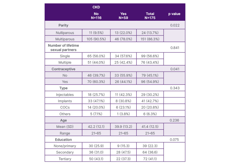
Table 1: Socio-demographic, clinical, and reproductive characteristics of women with and without CKD at MTRH
CKD: chronic kidney disease; COC: combined oral contraceptives; SD: standard deviation; MTRH: Moi Teaching and Referral Hospital.
The commonest abnormal cytology was atypical squamous cell of undetermined significance (ASCUS) at 5.1% overall. ASCUS was reported at 8.5% (5/59) among women with CKD and 3.4% (4/116) in women without CKD. The low-grade squamous intraepithelial lesions (LSIL) abnormal cytology accounted for 5.1% (3/59) in women with CKD and 1.7% (2/116) in women without CKD. High-grade squamous intraepithelial lesions (HSIL) were reported at 3.4% (2/59) among CKD women and 1.7% (2/116) among women without CKD (Table 2).
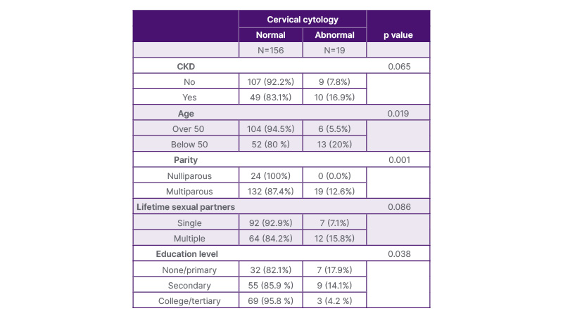
Table 2: Factors associated with abnormal cytology.
The mean age at CKD onset among those with abnormal cytology was 40.1 years versus 33.4 years for those with normal cytology (p=0.119). Diabetes was associated with more abnormal cytology (30.8%) than hypertension (10.2%), with statistical significance (p=0.008). Though systemic lupus erythematosus (33.3%) and chronic glomerulonephritis cases showed higher abnormal cytology rates, these were not statistically significant (p=0.266 and p=0.169, respectively).
Patients undergoing dialysis had more abnormal cytology (15.6%) compared to none among transplant recipients, with a significant association (p=0.002). Immunosuppressive regimens including tacrolimus, cyclosporine, mycophenolate mofetil, and steroids were not significantly associated with abnormal cytology (p values >0.3). One participant on prednisolone had abnormal cytology, but this too lacked significance (p=0.313; Table 3).
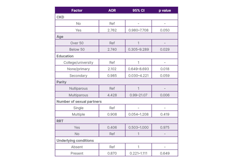
Table 3: Multivariate logistic regression analysis.
AOR: adjusted odds ratio; CKD: chronic kidney disease; RRT: renal replacement therapy.
Renal replacement therapy
Women with CKD were two times more likely to have abnormal cervical cytology compared to women without CKD, with a significant p value (AOR: 2.762; CI: 0.980–7.708; p=0.05). Factors found to be significantly associated with abnormal cervical cytology were age less than 50 years (AOR: 2.74; CI: 0.305–9.289; p=0.029). Women with no education or lower-level education, such as primary, were two times more likely to have abnormal cytology compared to women with college and secondary education (AOR: 2.012; CI: 0.649–8.693; p=0.018). Multiparous women were four times more likely to have abnormal cervical cytology than nulliparous women (AOR: 4.428; CI: 0.99–21.07; p=0.006).
DISCUSSION
This study evaluated cervical cytology through the use of Pap smears, as it remains a highly accessible and cost-effective screening tool. This allowed the study to reflect real-world conditions where molecular testing is not readily available while global public health is shifting towards the use of HPV detection due to its superior sensitivity. Australia is on track to eliminate cervical cancer as a public health problem by 2035 by implementing a dual approach through high HPV vaccination rate and HPV screening.
Prevalence of Abnormal Cervical Cytology
Cervical cancer remains a major public health issue in Kenya, causing 12.2% of all cancer cases and over 3,591 deaths in 2022.4 It accounts for about 3% of post-transplant malignancies, second only to skin cancer in female transplant recipients.8 A study in Denmark found that women with CKD or on dialysis had a higher likelihood of abnormal cytology than the general population.9
In this study, the prevalence of abnormal cervical cytology in women with CKD was 16.9%, compared to 7.8% in those without CKD. This trend may be linked to immunosuppression and chronic inflammation in CKD, impairing HPV clearance and increasing cytological abnormalities. A study at Kenyatta National Hospital, Nairobi, Kenya, reported a comparable prevalence of 12.5%,10 likely due to similar screening approaches and geographical setting. An Ohio study found a higher prevalence (27.5%), possibly due to more frequent screenings per protocol over time.11
Patterns of Abnormal Cytology
The most common abnormality in this study was ASCUS, found in 8.5% of CKD participants, followed by LSIL (5.1%), and HSIL (3.4%). In contrast, non-CKD women showed ASCUS (3.4%), LSIL (1.7%), HSIL (0.9%), and one case of microinvasive disease. This pattern mirrors findings in a USA study of transplant candidates, where ASCUS was most common.11 However, a study in Türkiye showed LSIL was more frequent, likely due to a younger participant age group, including adolescents more prone to transient HPV infections.12
Factors Associated with Abnormal Cytology
Women with CKD were significantly more likely to have abnormal cytology (AOR: 2.762; CI: 0.980–7.708; p=0.05), consistent with findings by Atılgan et al.,12 where patients with CKD had a threefold higher risk of preinvasive lesions due to immune dysfunction. Conversely, Haberal et al.13 found no such association, likely due to a smaller sample size.
Age below 50 was also associated with more abnormalities (AOR: 2.740; CI: 0.305–9.289; p=0.029), aligning with Chaung et al.’s11 study. However, Dalgaard et al.’s9 findings showed a higher risk among women aged 50–64, attributed to better health-seeking behaviours in a well-supported healthcare system.
Women with no or primary education had significantly more abnormalities (AOR: 2.012; CI: 0.649–8.693; p=0.018), a trend echoed in Zambia, where tertiary education reduced the risk of abnormal cytology.14 Low education may limit awareness and access to screening.
Multiparity was significantly associated with abnormal cytology (AOR: 4.428; CI: 0.99–21.07; p=0.006), likely due to increased HPV exposure and cervical trauma. This finding is supported by studies in Kenya and Indonesia, although contradicted by Bilgi et al.,16 who found no such link.10,15,16
Lastly, patients undergoing dialysis had more abnormalities (15.6%) than transplant recipients (0%), significant in bivariate but not multivariate analysis. This aligns with Kasiske et al.,17 who noted more cervical abnormalities in patients who underwent dialysis.
Limitations of the Study
This was a cross-sectional study, and therefore the cause and effects of the risk factors described could not be established. This was a single-centre study, and therefore the results can only be generalised to other similar-level facilities and not to the general population.
CONCLUSIONS
Women with CKD have a higher association of abnormal cervical cytology compared to women without CKD at MTRH. ASCUS was the commonest cytology pattern among women with CKD. Factors associated with abnormal cervical cytology were women having CKD, women below the age of 50 years, multiparous women, and women with no or primary education.
Increasing cervical cancer screening surveillance among women with CKD, multiparous women, women less than 50 years old, and women with no tertiary education, with frequent follow-up could reduce the high risk of abnormal cervical cytology among this group.