Abstract
Introduction: Uterine rupture is a rare, life-endangering complication in obstetrics, which is accompanied with high fetal and maternal morbidity and mortality, and occurs most commonly in women with a prior uterine scar, typically from a Caesarean section. However, the incidence of unscarred uterine rupture is extremely rare and poses unique diagnostic and management challenges due to its unpredictability and rapid clinical deterioration.
Case Report: Here is a case report on presentation and outcome of a case of rupture in a previously unscarred uterus in the third trimester. Placenta accreta spectrum, a recognised risk factor for uterine rupture, is uncommon in an unscarred uterus. This case has been considered for presentation due to its rarity, co-existence with placenta accreta spectrum, diagnostic dilemmas, and immediate surgical intervention, which led to the most favourable yet unexpected feto-maternal outcome evidenced by real-time photographs supplemented herewith.
Discussion: Risk factors for uterine rupture include high parity, uterotonic misuse, obstructed labour, uterine anomalies, connective tissue disorders, placenta accreta spectrum, and traumatic deliveries. Symptoms often manifest abruptly with sudden abdominal pain, fetal distress, vaginal bleeding, and signs of hypovolemic shock. Diagnosis may be clinical or assisted by imaging, though often confirmed intraoperatively. Perinatal mortality remains high and maternal morbidity includes haemorrhage, need for hysterectomy, and end organ damage.
Conclusion: This case report may throw some light on how high clinical suspicion, even in low-risk patients, is essential.
Key points
1. Uterine rupture and placenta accreta spectrum can occur even in an unscarred uterus, although it is a rare event, with significant risks to both mother and the fetus.2. In cases of spontaneous uterine rupture, early recognition and immediate surgical intervention are critical to improve maternal survival, reduce morbidity, and improve fetal outcome. In this case, it was observed that a timely intervention led to a successful live delivery followed by the recovery of the mother.
3. A high index of suspicion should be maintained, even in women without conventional risks factors such as previous Caesarean section or uterine surgery.
INTRODUCTION
Uterine rupture is a rare but serious emergency in obstetrics, typically associated with a scarred uterus from previous Caesarean delivery or other uterine surgeries like myomectomy.1 Unprovoked rupture of an unscarred uterus, particularly in the third trimester, is an exceptionally uncommon event, with an estimated incidence of one in 8,000 to one in 15,000 deliveries,2 with higher rates reported in low-resource settings.3 Despite its rarity, it carries significant risks of maternal haemorrhage, fetal compromise, and even mortality if not promptly diagnosed and managed.4
The clinical presentation is often non-specific, ranging from abdominal pain and abnormal fetal heart patterns to signs of hypovolemic shock,5 making early recognition challenging. Known risk factors include high parity, uterine overdistension, obstructed labour, misuse of uterotonics, and trauma;3,6 however, in some instances, no predisposing factors are identified. Most of the cases of uterine rupture are associated with fetal demise, and the rate can be as high as 50–75%, especially in unscarred uterine ruptures or in settings with delayed access to emergency care.4
This case report highlights a rare instance of placenta accreta spectrum causing rupture of an unscarred uterus in the third trimester, underscoring the imperative of maintaining vigilant clinical suspicion, even in low-risk patients, and emphasising the critical role of timely surgical intervention in optimising maternal outcome and delivery of a live fetus from the abdominal cavity.
CASE DESCRIPTION
A 40-year-old lady, G2P1A0L1, at 31 weeks 5 days of gestation, was admitted in the Department of Obstetrics and Gynecology after being referred from a peripheral hospital with complains of abdominal pain for 5 days in a suspected case of placenta previa. The pregnancy was spontaneously conceived and antenatal checkups had been uneventful. She had no history of previous Caesarean section, uterine surgery, trauma, or known uterine anomaly. There were also no significant medical or obstetric comorbidities. Ultrasonographies done during antenatal visits were not suggestive of any abnormal lie of the fetus.
On admission, the patient was haemodynamically stable with a BP of 120/70 mmHg, pulse rate of 88/min, and without any pallor. Abdominal examination revealed loss of uterine contour with no tenderness, and the fetal parts were palpable superficially. Fetal heart sound was present and regular, 144 bpm. An ultrasound revealed free fluid in the peritoneal cavity, suggestive of haemoperitoneum, and the presence of fetal cardiac activity in the abdominal cavity outside the uterus.
Given the clinical suspicion of uterine rupture, the patient was immediately taken up for emergency laparotomy under general anaesthesia. Intraoperatively, approximately 1 L of haemoperitoneum was noted. A complete rupture of the uterine wall was identified at the fundus of the uterus with the fetus entirely in the abdominal cavity and placenta densely adhered to the fundus of the uterus posteriorly, invading the myometrium with its lower end partially covering the internal os. A live male fetus weighing 1,880 grams was delivered from the left paracolic gutter (Figure 1). An attempt to separate the placenta failed, and an obstetric hysterectomy was done (Figure 2). The gross sample of uterus along with the placenta was sent for histopathological examination.
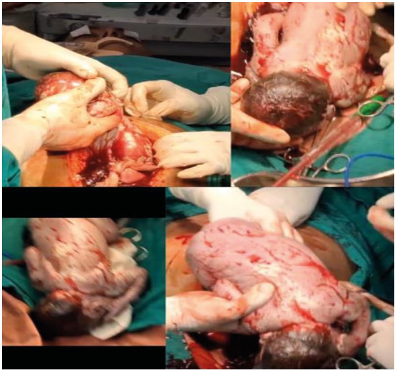
Figure 1: Delivery of a live baby from the left paracolic gutter.
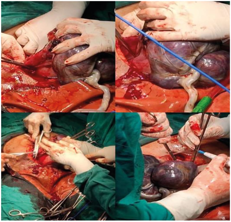
Figure 2: Obstetric subtotal hysterectomy.
The patient received two units of packed red blood cells and other supportive management postoperatively. She was monitored in the high dependency unit for a period of 7 days and she made an uneventful recovery. The baby was kept in the NICU for watchful observation and was free from any morbidity.
DISCUSSION
Uterine rupture is a critical obstetric emergency that typically occurs in the context of a previously scarred uterus, most commonly due to prior Caesarean section. However, rupture of an unscarred uterus is exceedingly rare, particularly in the third trimester, and presents significant diagnostic and management challenges. The present case highlights a spontaneous rupture in an unscarred uterus, in a preterm pregnancy with no identifiable risk factors, emphasising the unpredictable nature of this condition.
The incidence of unscarred uterine rupture is estimated at one in 8,000 to one in 15,000 pregnancies.2 The known risk factors include high parity, uterine anomalies, excessive use of uterotonic agents, obstructed labour, external trauma, and connective tissue disorders like Ehlers-Danlos syndrome.3,6 Nevertheless, cases without any predisposing factors, such as the one presented here, have been documented, raising concerns for idiopathic nature.
Clinical presentation is often subtle and non-specific, with symptoms such as abdominal pain, cessation of contractions, vaginal bleeding, or signs of hypovolemic shock.5 Loss of uterine contour, palpable fetal parts, and absence of fetal heart sounds are alarming signs. In this case, the patient complained of moderate abdominal pain, and a differential diagnosis of rupture was presumed after institutional ultrasonography, which was later confirmed intraoperatively.
Prompt recognition and immediate surgical intervention are vital to minimise adverse maternal and fetal outcomes. Intraoperative findings often include haemoperitoneum and complete uterine wall disruption, with the fetus possibly expelled into the peritoneal cavity. Uterine repair may be attempted if feasible, and if the patient is haemodynamically stable. In other instances, hysterectomy may be required,7 as in this case.
This case is unique in its presentation, as the uterine rupture happened at the fundus of an unscarred uterus with no specific symptoms of uterine rupture and without fetal demise. The histopathological examination revealed placenta previa with placenta increta. The rupture of the unscarred uterus could possibly be due to the invasion of the placenta into the myometrium (placenta accreta spectrum), leading to thinning of the myometrium and causing rupture. The survival of the fetus could possibly be justified due to the inseparable placenta densely adhered to the uterus, which probably maintained circulation and blood supply via the umbilical cord. Contrary to the age-old belief of 100% fetal demise in abdominal pregnancy secondary to ruptured uterus, a live baby was delivered from the peritoneal cavity, which was not only rare but also made the case reportable.
This case also underlines the importance of sustained clinical alertness for uterine rupture, even in women without typical risk factors. Early diagnosis, rapid resuscitation, and timely surgical management are the keys to favourable outcomes. Additionally, counselling regarding future pregnancies and delivery planning is essential, given the increased risk of recurrence.
Various cases of uterine rupture have been reported worldwide. Nevertheless, this presentation is exceptionally rare, with only few cases with similar combination of findings documented in the literature.
A nationwide population-based cohort study conducted in the Netherlands to assess the incidence of uterine rupture in scarred and unscarred uteri, and its maternal and fetal complications, concluded that 13% of the ruptures occurred in unscarred uteri and 72% occurred during spontaneous labour, and were associated with severe maternal and neonatal morbidity and mortality.8
In a study by Locher et al.,9 the sole potential risk factor was a closed adhesion of the small intestine to the left sacro-uterine ligament, indicating persistent inflammation. The authors found that lateral damage to an unscarred uterus increases the likelihood of maternal and fetal problems following rupture. Hence, for the patients presenting with unusual pain, cautious monitoring is important.
In another case report, a complete rupture of the posterior wall of the uterus with partial avulsion of the left ovary following induction of labour was noted. The patient underwent subtotal hysterectomy with left salpingectomy. The baby died in utero. The patient’s history of previous medical termination of pregnancies via curettage, resulting in an undiagnosed perforation, may have put her at higher risks. She also had induction of labour for her current pregnancy, which increased her risk.10
Another case was reported in which a primigravida with an unscarred uterus was admitted with preterm pre-labour membrane rupture at 36+4 weeks of gestation, abnormal fetal heart sounds, and sudden onset of severe abdomino-pelvic pain. Rupture happened before the initiation of regular uterine contractions and without any interventional oxytocin. Despite the immediate Caesarean birth, the newborn showed signs of severe acidosis. This example emphasises the significance of keeping a high level of suspicion for uterine rupture, even in the unusual scenario of a primigravida with an unscarred uterus. This case also demonstrates a possible link between prelabour rupture of membranes and placental abruption.11
Guèye et al.,12 concluded that the clinical indicators of uterine rupture during pregnancy can be misleading and ambiguous. It can be difficult to differentiate it from other abdominal problems such as pancreatitis, gallstones, and appendicitis. They also found that high parity was a significant risk factor for spontaneous uterine rupture.12
In another instance, a 34-year-old primigravid lady who was 31+3 weeks pregnant got an acute abdomen and pre-eclampsia. After an emergency laparotomy, the cause of the incident was determined to be a uterine rupture. There was a stillborn girl. The placenta’s pathological investigation revealed abruption, which may have contributed to this serious obstetrical problem, along with the pregnancy’s potential cornual or angular location.13
Miller et al.,14 in 1997, conducted a retrospective study to examine risk factors, as well as maternal and neonatal outcomes, in 10 cases of intrapartum rupture of unscarred uterus. They concluded that although intrapartum rupture of unscarred uterus is a rare obstetric emergency, the maternal and perinatal outcomes can be optimised by awareness of risk factors, recognition of clinical signs and symptoms, and prompt surgical intervention.
A study conducted by Al-Zirki et al.15 in Norway, to follow trends of uterine rupture over a period of 40 years, concluded that there was a sharply increasing trend of uterine rupture associated with obstetric interventions like augmentation with oxytocin, scarred uterus from a previous Caesarean section, and labour induction with prostaglandins or prostaglandins combined with oxytocin. However, the study could not explain the association entirely.15
CONCLUSION
Spontaneous rupture of an unscarred uterus in the third trimester is an extremely rare but potentially fatal obstetric emergency. Placenta accreta spectrum, although rare in unscarred uteri, can manifest as an important risk factor for rupture in such cases. This case highlights the need for clinicians to maintain a high degree of clinical suspicion, even in low-risk patients with no identifiable predisposing factors. Early recognition, prompt surgical intervention, and appropriate postoperative care are crucial in improving maternal outcomes. Contrary to the age-old idea of 100% fetal demise in secondary abdominal pregnancy, a live baby was delivered from the peritoneal cavity, which was not only rare but also made the case reportable. Furthermore, this case reinforces the importance of postpartum counselling and careful monitoring in subsequent pregnancies due to increased risk of recurrence in case of preservation of the uterus.