Meeting Summary
Chronic obstructive pulmonary disease (COPD) is a heterogeneous and progressive lung condition representing a leading cause of morbidity and mortality worldwide. Despite advances in COPD management, many patients continue to experience recurrent exacerbations and persistent disease burden.
At the American Thoracic Society (ATS) International Conference, Monica Kraft, Icahn School of Medicine at Mount Sinai, New York City, New York, USA, examined the biology of interleukin-33 (IL-33) and its role in the pathobiology of COPD. The symposium opened with an overview of the clinical burden of COPD, highlighting exacerbations as sentinel events associated with significant morbidity and mortality. The distinct but related manifestations of inflammation and mucus dysfunction were discussed, with lung function decline, exacerbation risk, and all-cause mortality highlighted for their clinical associations with mucus plugging.
Thereafter, Kraft discussed the role of IL-33 as an upstream alarmin cytokine, constitutively stored in epithelial cells, endothelial cells, and fibroblasts, and released in response to airway injury. IL-33 may drive COPD pathophysiology by affecting a variety of downstream cell types through two bioactive forms. The reduced form of IL-33 (IL-33 RED) signals via the serum stimulation-2 (ST2) receptor, driving heterogeneous Type 1, 2, and 3 airway inflammation. The oxidized form (IL-33 OX) signals independently via the receptor for advanced glycation end-products (RAGE)/epidermal growth factor receptor (EGFR) complex, promoting goblet cell hyperplasia, mucin hypersecretion, and impaired epithelial repair.
IL-33 expression is elevated in COPD and increasing levels correlate with more severe COPD. Elevated IL-33 in COPD has been associated with chronic bronchitis and exacerbation phenotypes, underscoring its relevance across the heterogeneous COPD population.
The Burden and Clinical Challenges of COPD
COPD is a heterogeneous lung condition characterized by chronic respiratory symptoms, including dyspnea, cough, sputum production, and/or exacerbations, resulting from abnormalities of the airways, alveoli, or both, which lead to persistent and often progressive airflow obstruction.1 COPD encompasses pathological changes such as bronchitis, bronchiolitis, and emphysema, reflecting the complex and multifactorial nature of the disease.1 Key hallmarks of COPD include recurrent exacerbations, mucus dysfunction, and abnormal inflammatory responses within the airways,1-5 all of which can contribute to disease progression, impaired lung function, and reduced quality of life.2,4
COPD remains a major global public health burden. In 2022, approximately 11.7 million people in the USA had been diagnosed with COPD, representing around 4.6% of the adult population.6 Furthermore, COPD is currently recognized as one of the top three leading causes of death worldwide,1 underscoring the substantial morbidity, mortality, and healthcare burden associated with the disease. Together, these data highlight the urgent need for improved disease management strategies and continued research into the underlying mechanisms and management of COPD.
Despite optimization of inhaled maintenance therapy, exacerbations remain a frequent and burdensome feature of COPD.1-3,7,8
Kraft opened the symposium presentation by describing data from the SIRIUS study, illustrating the scale of this ongoing clinical challenge. This USA retrospective cohort analysis identified 4,920 patients aged 40 years or older who were continuously receiving inhaled triple therapy, which included an inhaled corticosteroid, long-acting beta2-agonist, and long-acting muscarinic antagonist, and who had experienced at least two moderate or at least one severe exacerbation during a 12-month baseline period.7,8 Moderate exacerbations were defined as COPD-related emergency room, physician office, or outpatient visits accompanied by a prescription for an antibiotic or corticosteroid within ±7 days; severe exacerbations as hospitalizations of at least 2 days with a primary COPD diagnosis.7,8
At baseline, 89.6% (n=4,408) of patients had at least one prescription for oral corticosteroids, corresponding to a mean annual exposure of 73 days.7,8 During the first year of follow-up, 69% of patients experienced at least one further exacerbation of any severity, and 25% had at least one severe exacerbation, demonstrating that a substantial proportion of patients continued to experience exacerbations despite receiving triple therapy.7,8 Kraft described these exacerbations in COPD as a sentinel event associated with increased risks of mortality, underscoring current clinical challenges in this patient population.
Underlying this persistent exacerbation burden are two interrelated hallmarks of COPD pathophysiology that formed the central themes of the symposium: inflammation and mucus dysfunction. The most widely recognized mechanism, inflammation, stems from environmental insults and repeated epithelial damage that drive chronic airway inflammatory pathways and remodeling, worsening respiratory symptoms and increasing exacerbation risk.9,10 Additionally, mucus dysfunction, encompassing mucus hypersecretion, impaired airway clearance, and mucus plugging contribute to airway obstruction, structural damage, and further exacerbation risk.11,12 Kraft described this second mechanism as an increasingly recognized, distinct, but complementary process. Understanding the biological mechanisms that drive both processes is critical to understanding why patients may continue to deteriorate.
Mucus Dysfunction: Beyond Hypersecretion
Kraft described mucus dysfunction as central to the pathology of COPD. Mucus hypersecretion is associated with disrupted basal cell differentiation, excessive production of the mucin protein MUC5AC by goblet cells in the bronchial epithelium, and impaired ciliary clearance, favoring infection and microbial growth.13-16 These processes contribute to COPD symptoms including productive cough and dyspnea; productive cough may occur in up to 49% of patients with COPD, is linked to poorer clinical outcomes, and its prevalence often increases with disease severity.15,16 Distinct from hypersecretion, mucus plugs are viscous collections of mucus that obstruct the airway lumen and cause airway occlusion.5,17 Mucus plugs occluding the airways have been observed on CT scans in 25–67% of patients with COPD18,19 with prevalence increasing with Global Initiative for Chronic Obstructive Lung Disease (GOLD) disease stage.5 High mucus plug scores are associated with accelerated lung function decline20 and increased all-cause mortality.19 Kraft emphasized “when you see those [plugs] on a CT scan, it’s certainly worth noting and paying attention.”
The scale and heterogeneity of mucus dysfunction in COPD is further illustrated by data from the COPDGene study, an observational prospective cohort study of 45- to 80-year-old non-Hispanic White or non-Hispanic Black patients with a ≥10 pack-year smoking history.21 In a cross-sectional analysis of 4,363 participants with confirmed COPD (GOLD Stages 1–4), approximately 75% had at least one sign of mucus dysfunction, defined as cough, phlegm, and/or mucus plugs on CT. However, these manifestations were not uniform. Of those affected, approximately 45% had cough or phlegm only, without evidence of mucus plugging on CT; around 20% had silent mucus plugs only, with no associated cough or phlegm; and approximately 35% had both.21 Kraft explained that these findings underscore a critical distinction: mucus hypersecretion and mucus plugging are not synonymous, do not always co-exist, and likely reflect different, though potentially overlapping, pathological processes. The presence of silent plugs in a substantial proportion of patients serves as a particular reminder that mucus dysfunction may be present and clinically significant even in the absence of overt respiratory symptoms.21
The potential clinical consequences of mucus plugging extend across lung function, exacerbation risk, and mortality.5,20,22 Longitudinal data demonstrate that both persistent and newly formed mucus plugs are associated with accelerated decline in post-bronchodilator forced expiratory volume in 1 second (Post-BD FEV₁) over 5 years. Patients with persistently positive plugs experienced the greatest rate of decline at −60.4 mL/year, followed by those with newly formed plugs at −54.9 mL/year, nominally greater than those who were persistently plug-negative (−37.2 mL/year) or whose plugs resolved (−39.3 mL/year).22 The association with exacerbation risk was also emphasized. In a Korean hospital-based cohort of patients with COPD followed over 15 years, patients with mucus plugs faced a 2.1-fold greater risk of a severe COPD exacerbation (adjusted hazard ratio: 2.1; 95% CI: 1.43–3.10; p=0.001) and a 1.5-fold greater risk of a moderate-to-severe exacerbation (95% CI: 1.12–2.02) compared with those without plugs.20 The relationship between mucus plug burden and mortality is notably linear. In a separate USA-based analysis, all-cause mortality increased linearly with elevated mucus plug score, defined as the number of lung segments with mucus plugs. Compared to those with no mucus plugs, patients with a mucus plug score of 1–2 had an adjusted hazard ratio for all-cause mortality of 1.15 (95% CI: 1.02–1.29; p=0.02), rising to 1.24 (95% CI: 1.10–1.41; p<0.001) in those with a score of ≥3.5
Collectively, these data establish mucus plugging as a clinically significant manifestation of COPD, distinct from, though frequently coexistent with, mucus hypersecretion, and one that warrants greater recognition in both clinical practice and our understanding of the underlying disease biology.
IL-33 is an Upstream Alarmin Cytokine
Kraft introduced IL-33, an alarmin cytokine constitutively stored in the nuclei of structural barrier cells, including lung epithelial and endothelial cells and fibroblasts.23-26 Unlike cytokines that are actively secreted in response to immune stimulation, she described how IL-33 is released passively following necrosis and tissue injury, in response to cigarette smoke, pollutants, microbes, and other environmental stimuli, positioning it as an early danger signal at the site of airway damage.26
Increased IL-33 expression has been observed in both sputum and serum from patients with COPD relative to healthy controls, bringing attention to potentially meaningful underlying biology of IL-33 in COPD.27,28
Kraft explained that central to IL-33’s role in COPD is the airway epithelium, which acts not as an “innocent bystander but as an active participant” in immune regulation, functioning as an environmental sensor, driving innate and adaptive immune responses, and serving as a starting point for airway remodeling.29-31 Repeated epithelial injury and abnormal repair contribute to chronic airway inflammation and impaired epithelial integrity,13 which Kraft described is reflected histologically as marked goblet cell hyperplasia observed in mucosal biopsies from patients with COPD compared with healthy non-smokers.32 Kraft detailed that these structural changes are driven in part by alarmin upstream danger signals rapidly released from damaged epithelial cells to initiate and amplify the inflammatory cascade.30,33
Three alarmins have emerged as central drivers of airway inflammatory responses: IL-33, thymic stromal lymphopoietin (TSLP), and IL-25.30,33 Although all originate primarily from the airway epithelium, their cellular sources and biological reach differ.30,33 IL-33 is produced by both epithelial and endothelial cells, giving it a broader tissue distribution than TSLP, which is derived mainly from epithelial cells, or IL-25, which is predominantly produced by epithelial tuft chemosensory cells.30,33 Similar to TSLP, IL-33 acts on a broad range of immune and structural cells, whereas IL-25 has a comparatively narrower range of cellular targets.30
All three alarmins regulate immunity, induce downstream inflammatory pathways, and mediate potential airway remodeling and structural change.30 The disease associations of IL-33 and TSLP are similarly broad, with overexpression linked to asthma, atopic disease, COPD, bronchiectasis, lower respiratory tract disease, chronic rhinosinusitis, and eosinophilic esophagitis.30,33-40 Overexpression of IL-25 has also been linked to asthma, atopic disease, lower respiratory tract disease, and chronic rhinosinusitis.30,33-40
The inflammatory pathology of COPD is heterogeneous and reflects the interaction of multiple overlapping immune pathways triggered by epithelial injury from cigarette smoke, pollutants, allergens, bacteria, and viruses.10,26 Multiple downstream cell types are activated, most notably macrophages and neutrophils in Type 1 and Type 3 inflammatory responses and Group 2 innate lymphoid cells (ILC2), eosinophils, and mast cells in Type 2 inflammation.10,26 IL-33 and TSLP sit upstream of both Type 1/3 and Type 2 pathways, whereas IL-25 contributes predominantly to Type 2 responses.10,26 Kraft emphasized that heterogeneity in pathology is reflected across patients.
Focusing specifically on IL-33, Kraft then described how this cytokine may contribute to COPD pathophysiology through both inflammation and airway damage by affecting a variety of downstream cell types.41 IL-33 activates eosinophils, mast cells, and ILC2s involved in Type 2 inflammation, as well as macrophages, neutrophils, and endothelial cells associated with Type 1 and Type 3 responses.10,26,35,41 Beyond inflammation, IL-33 appears to promote epithelial remodeling associated with mucin hypersecretion and reduction in club cell and cilia-related genes.12 Human pulmonary vascular endothelial cells exposed to IL-33 in vitro produce IL-6, IL-8, and monocyte chemoattractant protein-1 (MCP-1), suggesting a potentially underrecognized role for endothelial activation in COPD pathobiology.37 IL-33 may also promote airway smooth muscle wound repair and fibroblast-driven extracellular matrix production, including collagen, fibronectin-1, and matrix metalloproteinases, collectively supporting a potential central role of IL-33 in chronic inflammation, airway remodeling, and progressive structural change in COPD.42-45
Exploring the Dual Roles of IL-33 in COPD Pathogenesis
The understanding of respiratory immunology has evolved considerably over recent decades, and with it, our appreciation of the role of IL-33 in airway disease. Kraft took the audience through a timeline of discoveries in respiratory immunology, starting with the first description of mast cells and eosinophils as far back as 1878–1879,46,47 to the discovery of IL-33 protein and mRNA in human endothelial cells in 2003,48 and the identification of IL-33 identified as an ST2-receptor ligand 2 years later in 2005.49 IL-33 was formally described as an alarmin in 2008,23 the same period in which ILC2s were characterized (2010),50 discoveries that collectively reshaped understanding of innate immune responses in the airway. The existence of an oxidized form of IL-33 (IL-33 OX) was first elucidated in 2015,51 but it was only more recently, in 2023, that IL-33 OX was identified as a bioactive agent in RAGE/EGFR signaling,12 a discovery that Kraft explained has significant implications for understanding the full breadth of the role of IL-33 in COPD pathobiology.
It is now recognized that both bioactive forms of IL-33 can contribute to the pathology of COPD in what Kraft described as “two sides to the coin” (Figure 1).10,12,26,35,51,52 Following airway epithelial damage, IL-33 RED is released from epithelial and endothelial cells, as well as fibroblasts, into the extracellular environment. Once released, IL-33 RED signals via the ST2 receptor, forming a complex with the IL-1 receptor accessory protein (IL-1RAP), driving both Type 1 and Type 3 inflammation, characterized by activation of macrophages, neutrophils, and endothelial cells, as well as Type 2 inflammation via eosinophils, mast cells, and ILC2s.10,26,35,52 Through this broad inflammatory activation, IL-33 RED can contribute to the heterogeneous airway inflammation characteristic of COPD.10,26,35,52 Critically, however, IL-33 RED is rapidly converted to its oxidized counterpart, IL-33 OX, in the extracellular environment.12,26,51 Unlike IL-33 RED, IL-33 OX does not signal via ST2; instead, it signals back to the epithelium and engages a distinct receptor complex comprising RAGE and EGFR.12 This ST2-independent pathway may drive a separate but complementary set of pathological consequences at the level of the airway epithelium, namely goblet cell hyperplasia, mucus hypersecretion, impaired epithelial repair, and impaired airway clearance.12,26,51 Together, these two pathways of IL-33 signaling, inflammation and mucus production, can converge to drive several hallmarks of COPD. Kraft emphasized that this combination may result in “significant airway injury,” and that understanding the clinical relevance of this biology requires consideration of how IL-33 is expressed in the airways of patients with the disease itself.
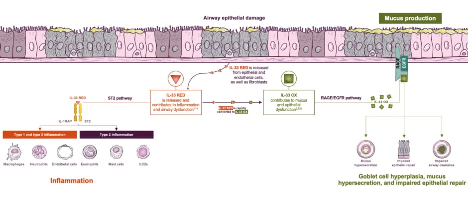
Figure 1: IL-33 RED drives Type 1, Type 2, and Type 3 inflammatory processes, while IL-33 OX contributes to
epithelial and mucus dysfunction.10,12,26,35,51,52
Please note that the proposed inflammatory pathways in COPD shown here have been simplified for illustration purposes only and do not imply clinical benefit or relevance.
EGFR: epidermal growth factor receptor; IL-1RAP: interleukin 1 receptor accessory protein; IL-33 OX: oxidized interleukin 33; IL-33 RED: reduced interleukin 33; ILC2: group 2 innate lymphoid cell; RAGE: receptor for advanced glycation end products; ST2: serum stimulation-2.
IL-33 Expression in COPD
Evidence from patient tissue and experimental airway models contributes to the clinical understanding of this dual IL-33 biology in COPD. Increased IL-33 expression has been demonstrated in the airways of patients with COPD,25 while a modeling study has found associations between IL-33 OX and epithelial abnormalities that characterize mucin hypersecretion and impaired epithelial repair.12 Kraft detailed lung homogenate analyses, which demonstrated that IL-33 levels were significantly higher in patients with GOLD Stage III/IV COPD compared with controls and further illustrated this with images of lung biopsies stained for IL-33 (Figure 2).25 Together, these data support an association between IL-33 and COPD.
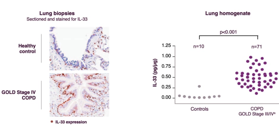
Figure 2: IL-33 levels tended to increase with COPD severity (GOLD stage).
aThis cohort comprised 17 GOLD Stage III and 54 patients with Stage IV COPD.
Lung biopsy images used with permission of Elsevier Inc, from Kearley J et al.;25 permission conveyed through Copyright Clearance Center, License No. [6207031273414].
COPD: chronic obstructive pulmonary disease; GOLD: Global Initiative for Chronic Obstructive Lung Disease.
The pathological associations of IL-33 OX on the airway epithelium have been demonstrated in bronchial air–liquid interface culture models.12 Immunohistochemistry of healthy bronchial air–liquid interface cultures following 7-day treatment with IL-33 OX (30 ng/mL) revealed a marked increase in MUC5AC/B-positive goblet cells compared with untreated controls, a pattern similar to that observed in COPD bronchial epithelial cultures, where goblet cell expansion is a well-recognized feature of the disease.12 Quantification of MUC5AC secreted into the apical region of these cultures found similar results.12 ELISA analysis demonstrated a statistically significant increase in MUC5AC secretion in IL-33 OX-treated cultures compared with that in untreated controls (p≤0.01), while cultures treated with IL-33 RED showed no significant difference from untreated controls.12 Kraft explained that the contrast between the two forms, with IL-33 OX but not IL-33 RED having associations with mucin hypersecretion, underscores the potentially distinct and complementary roles of each bioactive form, and highlights contributions from IL-33 OX signaling via the RAGE/EGFR pathway as a potential driver of the epithelial mucin hypersecretion phenotype observed in COPD.12
IL-33 OX has also been associated with impaired epithelial repair processes. In an epithelial scratch wound closure model using submerged cultures of growth factor-starved normal human bronchial epithelial cells, wound closure was assessed at 0 and 24 hours following treatment with IL-33 RED, IL-33 OX, or untreated control.12 At 24 hours, IL-33 OX-treated cultures displayed nominally lower percentage wound closure compared with both IL-33 RED-treated and untreated control cultures, suggesting that IL-33 OX may impair the capacity of the bronchial epithelium to repair itself following injury.12
Kraft highlighted that taken together, the MUC5AC hypersecretion and impaired wound closure findings point to an interesting observation: IL-33 OX, acting via the ST2-independent RAGE/EGFR pathway, could drive epithelial dysfunction similar to the pathological changes observed in COPD airways, including goblet cell hyperplasia, mucus hypersecretion, and failure of normal epithelial repair mechanisms.
Several studies have also investigated clinical features of patients with COPD, and found associations of higher levels of circulating IL-33 with both chronic bronchitis and exacerbations (Figure 3).27,53 In an analysis of 307 patients from the COPD Korean Obstructive Lung Disease cohort, patients with higher circulating IL-33 levels, defined as above the median cohort level of 11.9 pg/mL, had a significantly greater prevalence of chronic bronchitis compared with those with lower IL-33 levels (46.8% versus 32.7%; p=0.016).53 The association with exacerbation burden was similar: in a separate analysis of 62 patients with COPD, those in the upper quartile of IL-33 levels experienced a mean of 1.0 exacerbation per year compared with 0.4 in the lower IL-33 group (p<0.01), representing a 2.5-fold difference in exacerbation rate between the two groups.27
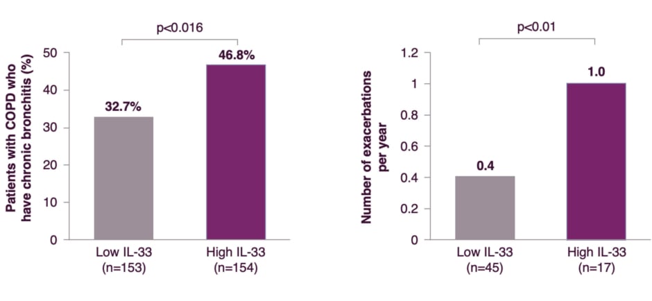
aThis study defined chronic bronchitis as phlegm for ≥3 months per year.53
bIn this analysis of 307 patients from the COPD Korean Obstructive Lung Disease cohort, IL-33 levels above the median IL-33 level of the cohort were defined as high, with all values below the median defined as low. At baseline, the median IL-33 level was 11.9 pg/mL (interquartile range: 7.9–30.6).53
cIn this analysis of 62 patients with COPD based in Korea, levels of IL-33 in the upper quartile of the cohort were
defined as high, with all levels below this value defined as low. Patients were prospectively followed for 1 year and monitored for exacerbation.27
dNumber of exacerbations per year (±SD): Low IL-33 group = 0.40 (±0.62) and High IL-33 group = 1.00 (±1.16).27
Kraft summarized that these clinical findings reinforce the pathobiological evidence, that IL-33 dysregulation may not only be a feature of COPD airways at the molecular and cellular level but has also been associated with clinical manifestations that drive disease burden: chronic bronchitis, mucus dysfunction, and recurrent exacerbations, across the heterogeneous COPD population.
Conclusion
IL-33 is an upstream alarmin cytokine with a broad and clinically relevant role in COPD pathobiology. Through its reduced form, IL-33 RED binds to the ST2 receptor to initiate and amplify heterogeneous Type 1, Type 2, and Type 3 airway inflammation. Through its oxidized form, IL-33 OX engages a distinct RAGE/EGFR signaling complex which can contribute to goblet cell hyperplasia, mucin hypersecretion, and impaired epithelial repair, consequences that closely mirror airway changes observed in COPD. Clinically, elevated IL-33 has been associated with chronic bronchitis, greater exacerbation frequency, and more severe disease.
Together, the dual pathways of IL-33 signaling may perpetuate a cycle of airway injury and dysfunction central to COPD pathobiology. As understanding of respiratory immunology has continued to evolve, the science behind IL-33 has become of increasing interest for clinicians managing this heterogeneous and burdensome disease.
US-113599 Last Updated 7/26