Abstract
Idiopathic chondrolysis of the hip (ICH) joint is rare after skeletal maturity. This study aimed to describe two cases of ICH in adolescent girls after skeletal maturity. The authors conducted a retrospective analysis of the clinical features, investigations, and management of two adolescent girls with ICH: a 16-year-old with bilateral hip involvement and an 18-year-old with unilateral hip involvement. Both of them presented with pain and limitation of hip movement. There was apparent lengthening, pelvic obliquity, and muscle wasting. The X-ray showed severe narrowing of the joint space consistent with ICH. This was managed with traction, analgesics, and a short course of steroids. A subtotal capsulectomy was subsequently performed. Both patients experienced symptom relief and an increase in the range of movement. In conclusion, ICH is diagnosed by excluding other causes of painful hips in children. It has characteristic clinical and radiological features. Traction and analgesics can provide short-term pain relief. Subtotal capsulectomy can improve the range of movement.
Key Points
1. Idiopathic chondrolysis is a diagnosis of exclusion in a teenage girl with a painful limp.2. This report describes two cases of idiopathic chondrolysis in adolescent girls after skeletal maturity.
3. Subtotal capsulectomy can improve hip range of motion and delay definitive procedures like total hip replacement.
INTRODUCTION
Idiopathic chondrolysis of the hip (ICH) is a rare musculoskeletal disorder characterized by accelerated destruction of articular cartilage, followed by remodeling and degeneration of the hip joint. Common symptoms include pain and limping. The condition predominantly affects teenage girls and shows a higher prevalence in African and Asian populations. The etiology of ICH remains unclear. Although anecdotal reports of complete recovery exist, the majority of cases progress to fibrous ankylosis or severe hip joint degeneration.1
ICH is uncommon after skeletal maturity. This article presents the clinical features and treatment of two cases of ICH in skeletally mature adolescent girls.
CASE PRESENTATIONS
The authors examined the medical records of two girls with ICH, and a retrospective analysis of their clinical features, investigations, management, and follow-ups was done.
Case 1
A 16-year-old girl presented with a 1-year history of bilateral hip pain, initially affecting the right hip. She received traditional (Ayurvedic) medications, which provided minimal relief. After approximately 4 months, pain developed in the left hip as well. The pain progressively worsened, resulting in significant functional impairment and inability to perform daily activities. There was no history of trauma or constitutional symptoms. The patient was unable to walk more than 500 meters, sit cross-legged, squat, or attend school.
At presentation, the patient exhibited an antalgic gait and required a walker for ambulation. Bilateral hip tenderness was noted. Exaggerated lumbar lordosis was present, attributed to hip flexion deformity. The right anterior superior iliac spine was positioned lower, consistent with an abduction deformity. Both hips demonstrated external rotation deformities. Range of motion was severely restricted in all directions. The right hip showed 30 degrees of flexion deformity with an additional 10 degrees of flexion possible, 15 degrees of abduction, and an external rotation deformity with no further movement. The left hip demonstrated 15 degrees of flexion deformity with an additional 10 degrees of flexion. Abduction deformity was 10 degrees with no further abduction. The limb was in neutral rotation with no further internal or external rotation possible.
Routine blood investigations demonstrated a hemoglobin level of 11 g/dL and a total leukocyte count of 5,300 cells/µL. The erythrocyte sedimentation rate was 15 mm/hr, and C-reactive protein (CRP) was 1.2 mg/L. The Mantoux test was negative. Serum rheumatoid factor, antistreptolysin O, and antinuclear antibody levels were within normal limits. Plain radiography revealed periarticular osteopenia, severe loss of articular cartilage, and global narrowing of the joint space bilaterally. Protrusio acetabuli was observed on the right side (Figure 1)
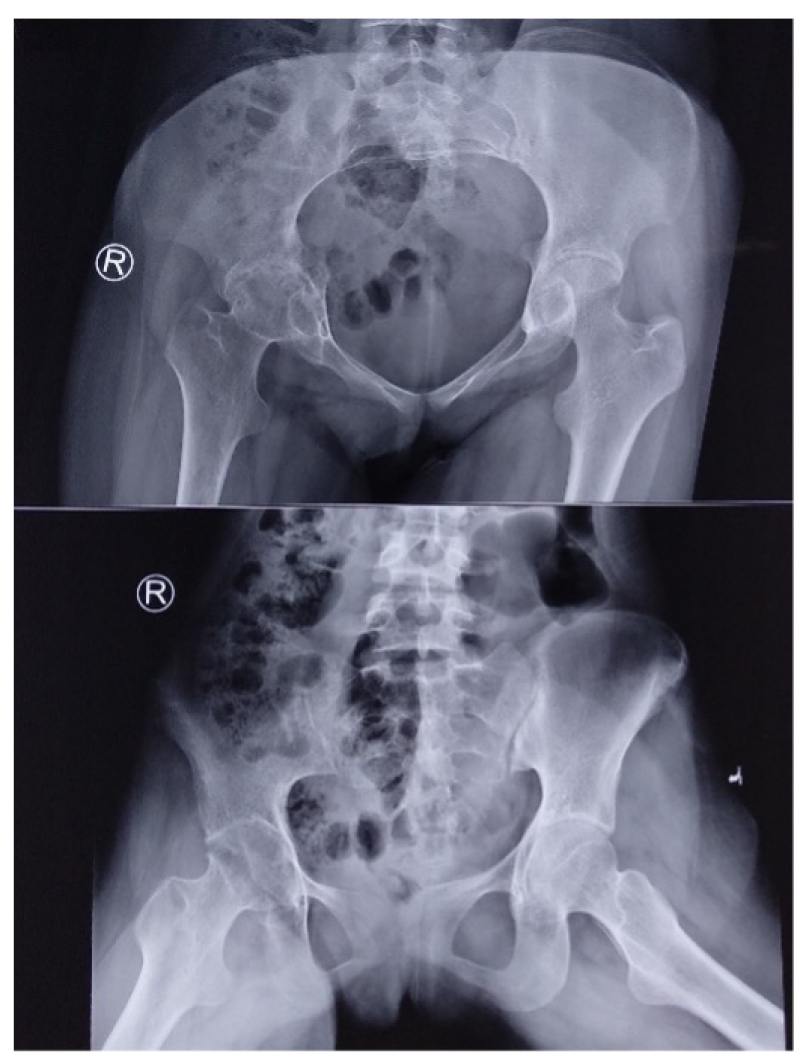
Figure 1: Radiographs of case 1.
Plain radiograph of the pelvis with both hips anteroposterior and lateral view showing periarticular osteopenia, concentric narrowing of the joint space, loss of subchondral sclerosis, and protucioacetabuli on the right hip; and periarticular osteopenia, narrowing of the joint space, and early protrucio on the left.
Aspiration of the joint yielded pale yellow synovial fluid with a total leukocyte count of 65 cells/µL, less than 10% neutrophils, synovial fluid glucose of 10 mg/dL, and protein concentration of 2 g/dL. Synovial fluid culture was negative, and a cartridge-based nucleic acid amplification test (CBNAAT) for tubercular bacteria was also negative.
Based on clinical and investigative findings, the patient was diagnosed with idiopathic chondrolysis of both hips. She was treated with bilateral above-knee skin traction for 3 weeks. Naproxen 250 mg was administered twice daily for pain relief, and a short course of oral methylprednisolone was provided for 1 week. Her pain subsided and her walking distance improved; however, she remained unable to squat or sit cross-legged. Similar treatment was administered four times within 1 year, but symptomatic improvement was not sustained. Consequently, subtotal capsulectomy of both hips was performed. Following capsulectomy, the patient achieved 90 degrees of flexion, 20 degrees of abduction, and approximately 15 degrees of both internal and external rotation in the right hip. On the left side, 110 degrees of flexion, 30 degrees of abduction, and 20 degrees of free rotation were observed. She was able to perform activities of daily living and return to school. At the 36-month follow-up, she remained asymptomatic.
Case 2
An 18-year-old female presented with a 6-month history of left hip pain and difficulty walking. She had received intermittent analgesic treatment during this period. There was no history of trauma or constitutional symptoms. Examination revealed an apparent lengthening of 2 cm in the left lower limb, pelvic obliquity with the left pelvis at a lower level, and a flexion deformity of 30 degrees with further flexion possible up to 80 degrees. The left hip was fixed in 20 degrees of abduction and 20 degrees of external rotation deformity. The patient was ambulant with a painful, fixed abduction gait.
Routine blood investigations revealed a hemoglobin level of 12 g/dL and a total leukocyte count of 4,320 cells/µL. The erythrocyte sedimentation rate was 8 mm/hr, and CRP was 0.8 mg/L. The Mantoux test was negative. Additional investigations to exclude infectious or rheumatologic etiologies were also negative. Plain radiography demonstrated severe pelvic obliquity, periarticular osteoporosis, loss of subchondral sclerosis, and marked concentric reduction of the joint space (Figures 2A and 2B). MRI revealed extensive thinning and destruction of the articular cartilage, subchondral cortical irregularity, and bone marrow edema involving the femoral head, acetabulum, neck, and intertrochanteric region. Minimal joint effusion was present, with no periarticular collection observed (Figure 2C).
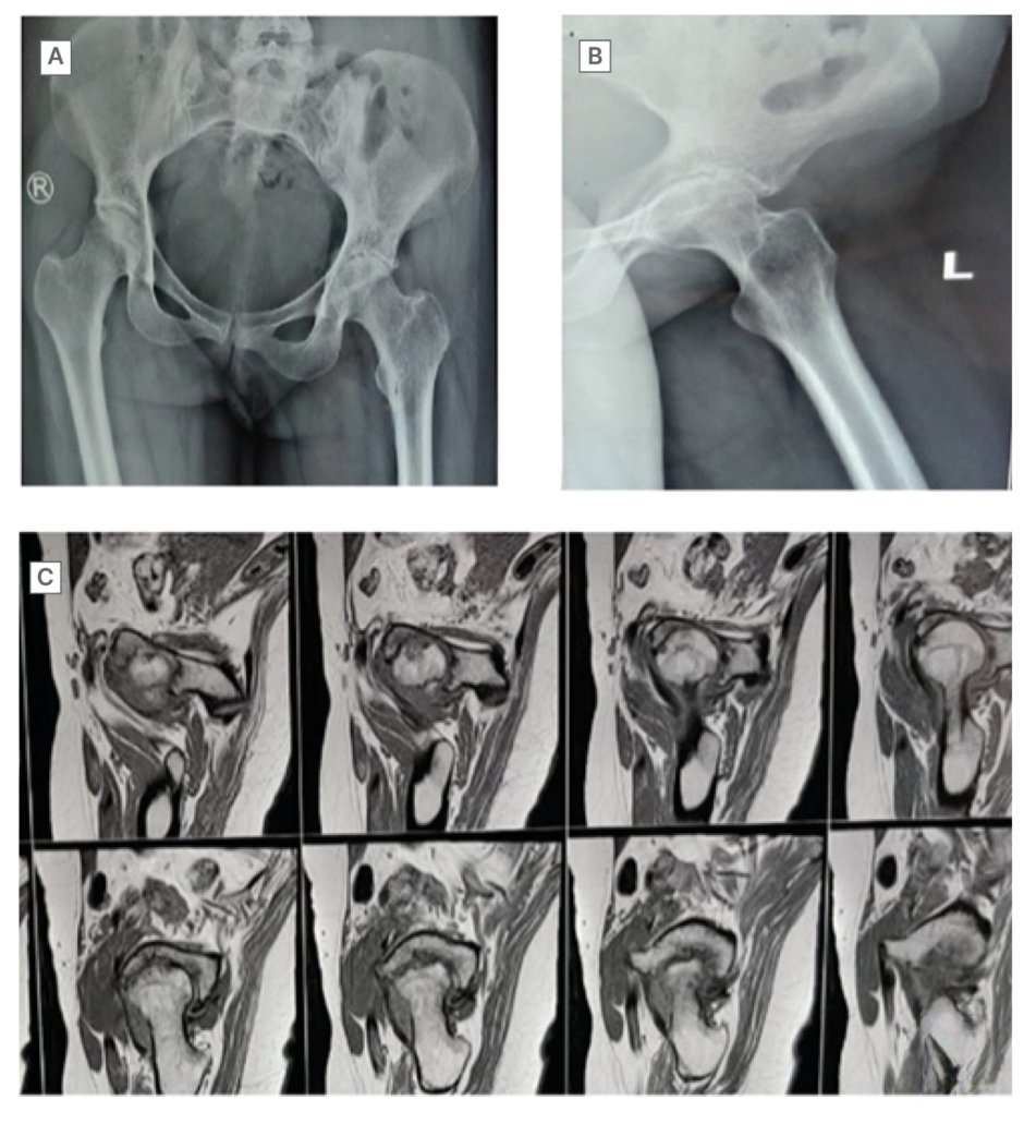
Figure 2: Imaging features of case 2.
A) Plain X-ray of the pelvis with both hips in the anteroposterior view showing periarticular osteopenia, global narrowing of the joint space, and an abduction deformity with pelvic obliquity on the left hip joint. B) Lateral view of the left hip showing osteopenia and narrowing of the joint space. C) MRI scan of the left hip showing extensive thinning and destruction of the articular cartilage. There is subchondral cortical irregularity and subchondral bone marrow edema of the femoral head and acetabulum. Bone marrow edema of the head, neck, and intertrochanteric region.
Synovial fluid analysis showed a pale-yellow color, normal cell count, and no elevation in protein or reduction in glucose levels. CBNAAT and culture of the synovial fluid were negative for tuberculosis and bacterial infections. After exclusion of other conditions, a diagnosis of ICH was established.
The patient received above-knee skin traction, analgesics, and a short course of steroids. Although her pain decreased, hip stiffness and deformity persisted. After approximately 6 months of non-operative management, a subtotal capsulectomy was performed. Following surgery, her pain resolved and her range of motion improved. She achieved a near-normal range of flexion and abduction, with approximately 30 degrees of internal and external rotation. At the most recent follow-up, 26 months postoperative, she continued to do well.
SURGICAL TREATMENT
The authors performed a standard subtotal capsulectomy in both cases. Pre-operative antibiotic prophylaxis consisted of 1.5 g cefuroxime administered 30 minutes before incision and continued twice daily for 48 hours. The procedure was conducted under a subarachnoid block. Each patient was positioned supine with a wedge placed beneath the ipsilateral buttock. The Smith Peterson approach was utilized. The abductors were elevated from the ilium, and the tensor fascia lata was released to address the abduction deformity. The straight head of the rectus femoris was released, and a tenotomy of the psoas muscle was performed to reduce the flexion contracture. A 1 cm strip of the capsule was excised along the entire arc of the hip joint, except for the posterior 90 degrees, to preserve the blood supply. Biopsies were obtained from the synovium and the periphery of the femoral head, and the specimens were sent for histopathology and culture. The wound was closed in layers after achieving hemostasis, with a suction drain left in situ.
Postoperatively, the drain was removed after 24 hours. Bilateral skin traction with a 3 kg weight was applied for 10 days. Sutures were removed on the tenth day. Synovial culture results were negative for bacteria and tuberculosis. Histopathological examination did not indicate infective or inflammatory conditions. CBNAAT of synovial tissue was negative for tuberculosis for both patients. Following suture removal, physiotherapy commenced, and active mobilization using a continuous passive motion device was initiated. Mobilization was maintained for 6 weeks.
DISCUSSION
ICH is a rare musculoskeletal disorder in children. Waldenström first described chondrolysis of the hip as acute necrosis of cartilage in the slipped capital femoral epiphysis.2 Trauma, infection, and prolonged immobilization can lead to chondrolysis of the hip. Duncan coined the term idiopathic chondrolysis of the hip in children where chondrolysis occurred without any predisposing causes. Rapidly progressive destruction of the articular cartilage is the cardinal finding.3 It can lead to early degeneration of the joint and secondary osteoarthritis.4 The female-to-male incidence ratio is 6:1. The right side is involved in most cases. Bilateral involvement and occurrence in adults are rare.5 The exact incidence of this disease is unknown, though there is a high incidence of severe cases in Black and South Indian populations.6 The literature on ICH is scarce, and most available literature includes anecdotal series and case reports. There can be difficulty in diagnosing ICH because of similarities in its presentation with some forms of juvenile idiopathic arthritis.7
There are many theories regarding the origin of ICH. Repetitive microtrauma, acute minor trauma, bacterial infection, and autoimmune reactions are some of the proposed mechanisms. Another theory suggests that increased intracapsular pressure leads to ischemia and cartilage loss. Nutritional deficiencies and endocrine and genetic factors can cause increased intracapsular pressure.8 Articular cartilage destruction by an autoimmune process in genetically susceptible individuals is the most widely accepted theory. The infiltration of inflammatory cells, including plasma cells, into the synovium supports this theory. The chondrolytic enzymes released during trauma or an elevated intracapsular pressure due to hemarthrosis are the basis for the mechanical theory for the origin of ICH. There are no similarities between the pathological features of cartilage destruction in idiopathic chondrolysis and osteoarthritis.9
Most cases are reported in teenage girls in Asia and Africa. ICH is monoarticular in 50% of patients. Gradual onset of pain and restriction of hip movements are common presentations. There is severe stiffness in the hip–antalgic gait at presentation. Flexion, abduction, and external rotation contractures develop at later stages. The anterior superior iliac spine on the affected side is at a lower level. There is apparent lengthening of the affected limb. There is a wasting of the gluteal and thigh muscles. There are no predisposing factors in most patients, though some reports suggest a history of trauma before the onset of symptoms.10 Septic arthritis, juvenile chronic arthritis, trauma, slipped capital femoral epiphysis, and Perthes disease can cause secondary chondrolysis in the hip. ICH is diagnosed by the exclusion of these secondary causes.11 The natural history of ICH has evolved over many years. The acute phase lasts for approximately 18 months. In this phase, there is inflammation of the synovium of the hip joint with progressive loss of articular cartilage of the femoral head and acetabulum. There is a progressive loss of movement of the affected hip. Later, the inflammation decreases and enters a chronic phase lasting approximately 3–5 years. In this phase, there are three possible outcomes. In the first group, cartilage replenishes, leading to a return to the normal range of movement and complete resolution of symptoms. The second group of patients develop a painless, stiff, ankylosed hip. The last group deteriorates to painful ankylosis in malposition.12
Routine blood tests, including total and differential leukocyte counts, are normal, with marginal elevation of the erythrocyte sedimentation rate in some cases. Serum levels of CRP, rheumatoid factor, antinuclear antibodies, and culture of the joint aspirate are normal in patients with ICH.13 The plain radiographs show some classical findings. There is periarticular osteoporosis and circumferential narrowing of the joint space to less than 1 mm. In some cases, the absence of a subchondral bone line, premature growth plate closure, and acetabulum protrusion is seen. There can be overgrowth of the femoral head and neck if radiography is performed several weeks to months after the onset of symptoms. MRI can help differentiate it from secondary causes and other conditions, such as tuberculosis, reflex sympathetic dystrophy, and pigmented villous nodular synovitis.14 In the early stages, the MRI scan shows a geometric focal area of bone marrow edema in the middle third of the proximal femoral epiphysis extending peripherally into the articular cartilage. There is no joint effusion, mild to moderate synovial enhancement, and minimal loss of articular cartilage. In advanced cases, there is severe loss of central cartilage in the femoral head and bone marrow edema involving the femoral head, neck, and acetabulum. There is no joint effusion or synovial thickening, and marked wasting of the gluteal and abductor muscles on the affected side. Bone scintigraphy revealed a diffuse increase in uptake in the periarticular region, suggesting an inflammatory reaction. Bone scans can detect abnormalities earlier than other investigations. A CT scan can provide more information than a plain radiograph about cartilage destruction, but its routine use is not advised due to the heavy dose of radiation for children.15-18 Histopathology examination reveals synovium hyperplasia with loss of articular cartilage and superficial cartilage necrosis. There is infiltration by inflammatory cells, including lymphocytes and plasma cells, and varying degrees of fibrosis.19
There are many differential diagnoses for ICH. Any condition causing a painful limp in a child can be a differential diagnosis. Most pediatric hip conditions are associated with adduction contracture of the hip. ICH, transient synovitis of the hip, occasionally tuberculosis, and juvenile chronic arthritis can cause abduction deformity of the hip in children. Transient synovitis is a self-limiting condition characterized by painful limping. A viral or bacterial infection precedes it, and there are no radiological abnormalities. Tuberculosis of the hip can produce abduction deformity when the ligament of Bigelow is destroyed due to posture and in the wandering acetabulum. Radiologically, there is juxta-articular erosion, concentric narrowing of the joint space, and periarticular osteopenia, similar to ICH. Therefore, tuberculosis of the hip needs to be ruled out before diagnosing ICH, especially in endemic regions.20 Isolated involvement of the hip is rare in juvenile patients with chronic arthritis. Serological markers and extra-articular manifestations can help
diagnose tuberculosis.1,9
Studies are scarce about ICH. The majority are isolated reports or small case series (Table 1).
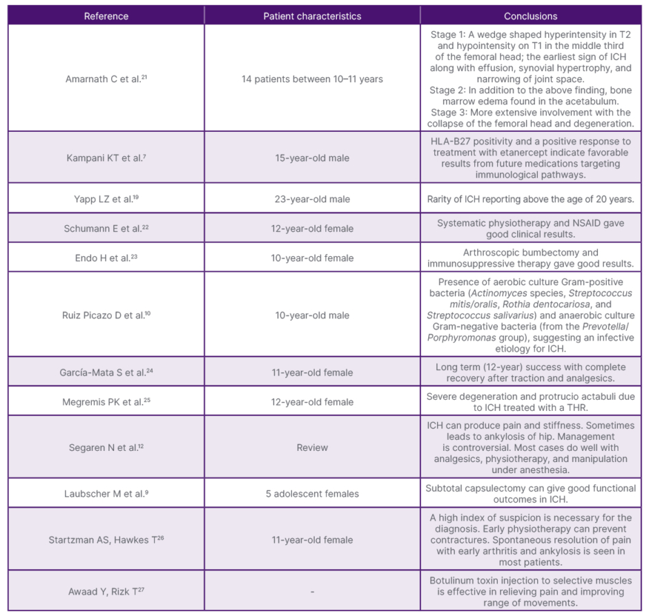
Table 1: Details of a few previously published cases with their conclusions.
ICH: idiopathic chondrolysis of the hip; NSAID: nonsteroidal anti-inflammatory drugs; T1: T1-weighted MRI; T2: T2-weighted MRI; THR: total hip replacement.
Various treatment modalities were tried for ICH, but none have been proven to be 100% successful: nonsteroidal anti-inflammatory drugs, steroids, physical therapy, traction, and etanercept are some of them. Subtotal capsulectomy and mobilization were attempted. There is a report of total hip replacement performed in a patient with ICH. Most non-operative treatments lead to disease recurrence and rapid progression to degenerative arthritis.1
At present, etanercept/TNF-α inhibition is the most notable emerging pharmacological approach specifically reported for idiopathic chondrolysis, but evidence remains limited to case-level data. There is no proven disease-modifying drug for ICH, and contemporary management remains predominantly multimodal, combining anti-inflammatory treatment, maintenance of motion, and appropriately selected surgical intervention.28
Recent surgical strategies for ICH increasingly emphasize joint preservation, especially during early disease stages. Hip arthroscopy with synovectomy or debridement, labral treatment, abrasion chondroplasty, and management of secondary femoral head deformity, such as arthroscopic osteochondroplasty, have been reported in selected cases and may contribute to motion preservation. Arthroscopic or open capsular release or capsulectomy is recommended when severe capsular contracture and stiffness are predominant. Joint distraction or arthrodiastasis represents an additional joint-preserving approach designed to unload articular cartilage and facilitate potential cartilage recovery, although supporting evidence remains limited. In advanced disease characterized by extensive cartilage loss or painful ankylosis, total hip arthroplasty has increasingly supplanted historical procedures, including excision arthroplasty and arthrodesis, particularly in skeletally mature patients. Nevertheless, due to the rarity of ICH and the predominance of case reports and small series in the literature, no surgical technique has demonstrated clear superiority.23,29,30
There are very few reports of ICH occurring after skeletal maturity. The presentation and progression of the disease are similar to those that occur before skeletal maturity.5,19,31
These two cases demonstrated that ICH can occur in adolescent girls even after skeletal maturity. Non-operative management can result in a short-term improvement of symptoms after skeletal maturity. Subtotal capsulectomy can decrease stiffness and increase the range of movements. The
long-term outcomes are unknown.
The following conclusions are from the experience of two cases of ICH occurring after skeletal maturity. Long-term outcomes and treatment algorithms for such patients can be available with studies involving more patients in the future.
CONCLUSION
Idiopathic chondrolysis of the hip after skeletal maturity is very rare. The diagnosis is based on clinical and radiological features and by excluding similar conditions. Traction and analgesics can give short-term relief from pain. Subtotal capsulectomy can increase the range of movements.