Abstract
Vacuoles, E1 enzyme, X-linked, autoinflammatory, somatic (VEXAS) syndrome is an adult-onset autoinflammatory disorder caused by somatic UBA1 mutations that predominantly affects men older than 50 years, presenting with recurrent fever, neutrophilic dermatoses, chondritis, pulmonary involvement, and macrocytic anemia. Distinguishing autoinflammatory flares from superimposed infection remains a major clinical challenge, particularly in patients receiving chronic immunosuppression. This case report describes a 76-year-old man with genetically confirmed VEXAS syndrome who presented with high-grade fever and a single blood culture positive for Actinomyces species. Despite empiric antibiotics, he defervesced rapidly, repeat blood cultures and imaging were negative, and his overall course was interpreted as more consistent with an autoinflammatory flare than with true infection, although concurrent glucocorticoid escalation and Actinomyces-active antimicrobial therapy limit definitive attribution. Several features supported this interpretation, including a history of recurrent unexplained fever on prednisone tapering and isolation of Actinomyces neuii from only one of two blood culture sets. The absence of leukocytosis and the borderline procalcitonin elevation are difficult to interpret given concurrent JAK inhibition, which can blunt these markers independent of infection. This case illustrates the diagnostic overlap between infection and autoinflammation in VEXAS syndrome and highlights the limitations of current biomarkers, including C-reactive protein (CRP) and procalcitonin, in distinguishing the two. Improved diagnostic tools and validated response criteria are needed to guide antibiotic and immunosuppressive management in this patient population.
Key Points
1.Recurrent fever is a core manifestation of vacuoles, E1 enzyme, X-linked, autoinflammatory, somatic (VEXAS) syndrome, an adult-onset autoinflammatory condition, yet affected patients also carry an intrinsic immune deficiency that predisposes them to serious infections. Reliably distinguishing an autoinflammatory flare from true infection is therefore a recurring and high-stakes diagnostic challenge in this population.2.This article describes a patient with genetically confirmed VEXAS syndrome who developed high-grade fever and a single blood culture positive for Actinomyces species. Despite empiric antibiotics, he defervesced rapidly, repeat cultures and imaging were negative, and his overall course was interpreted as more consistent with an autoinflammatory flare than with true bacteremia, although this attribution is confounded by concurrent glucocorticoid escalation and Actinomyces-active antimicrobial therapy.
3.Clinicians should be cautious about attributing fever solely to infection in VEXAS syndrome: absence of leukocytosis, a single positive blood culture set, and rapid clinical improvement may favor autoinflammation, but inflammatory markers can be blunted by JAK inhibition, and defervescence occurred while an Actinomyces-active antibiotic and higher-dose glucocorticoids were both being given. Improved biomarkers and validated response criteria are needed to guide antibiotic and immunosuppressive decisions.
INTRODUCTION
Vacuoles, E1 enzyme, X-linked, autoinflammatory, somatic (VEXAS) syndrome is an adult-onset autoinflammatory disorder caused by somatic UBA1 mutations, predominantly affecting men older than 50 years.1-3 The syndrome is characterized by recurrent fever, neutrophilic skin lesions, chondritis, lung involvement, macrocytic anemia, and vacuolization of myeloid and erythroid precursors within the bone marrow.1,3,4 Distinguishing disease-related inflammatory flares from superimposed infections is a major ongoing clinical challenge, particularly for patients receiving chronic immunosuppression. This report presents a patient with genetically confirmed VEXAS syndrome who developed high-grade fever together with a single positive blood culture for Actinomyces, illustrating this diagnostic uncertainty.
CASE DESCRIPTION
The patient was a 76-year-old man with VEXAS syndrome, chronic macrocytic anemia, and inflammatory polyarthralgia who presented with fever at 40.6 °C (105 °F) without localizing symptoms. He was diagnosed with VEXAS syndrome after extensive rheumatologic evaluation for persistent polyarthralgia, synovitis, and diffuse maculopapular rash. Skin biopsy and genetic testing revealed pathogenic UBA1 mutation (p.Met41Leu, variant allele frequency 43.7%). Bone marrow biopsy showed hypercellular marrow with trilineage hematopoiesis and cytoplasmic vacuolization of myeloid and erythroid precursors without dysplasia. He was receiving prednisone 20 mg daily and tofacitinib 5 mg once daily at the time of presentation; monthly tocilizumab had been under consideration in the months before this admission, but had not yet been initiated. Notably, the patient had a history of recurrent non-infectious fevers when his prednisone was tapered below 20 mg. He was not receiving anti-infective prophylaxis at the time of this admission.
On presentation, the patient was tachycardic but hemodynamically stable. Laboratory testing revealed a white blood cell count of 4.5×10⁹ /L, with neutrophils at 40.7%, lymphocytes at 45%, monocytes at 10.9%, eosinophils at 0.1%, hemoglobin of 8.5 g/dL, mean corpuscular volume of 109.2 fL, platelet count of 80,000/μL, erythrocyte sedimentation rate of 36 mm/hr, C-reactive protein (CRP) of 8.3 mg/dL, and procalcitonin of 0.52 ng/mL. Liver enzyme levels were normal. Urinalysis, respiratory viral testing, and chest radiography findings were unremarkable. One of two blood culture sets (one of four bottles) grew an anaerobic gram-positive rod, subsequently identified as Actinomyces neuii. Empiric vancomycin and ampicillin-sulbactam were initiated, and vancomycin was later discontinued because of a low suspicion of methicillin-resistant Staphylococcus aureus infection. Prednisone and tofacitinib were initially held on admission. CT of the abdomen and pelvis, along with abdominal ultrasonography, transthoracic echocardiography, and venous duplex studies, yielded negative results. Tests for tick-borne and viral infections during the recent admission were negative. Two additional sets of blood cultures, obtained after antimicrobial therapy had already been started, were negative, and the patient remained afebrile for the rest of the hospitalization. Rheumatology was consulted and recommended increasing prednisone to 40 mg, suspecting an autoinflammatory flare. His white blood cell count declined from baseline to 3.4×10⁹ /L, with neutrophils at 76.9%, lymphocytes at 7.9%, and monocytes at 8.2%, while hemoglobin dropped to 7.9 g/dL during the hospital stay. A review of prior laboratory trends revealed longstanding anemia and thrombocytopenia for several years, consistent with known VEXAS-related hematologic involvement. He was discharged on oral amoxicillin to complete a 10-day course and a prednisone taper.
Over the following 6 months, he was admitted to the hospital multiple times with a similar clinical picture, each time stabilizing on a prednisone taper. His outpatient regimen was changed from tofacitinib to intravenous tocilizumab, which was discontinued after an infusion reaction, and then to subcutaneous anakinra 100 mg daily approximately 1 month after the index admission. Following this switch, he had an initial period of good disease control, with no high fevers or skin flares reported on outpatient follow-up. He was subsequently admitted five more times over the following several months for fever in the pattern of his VEXAS flares. Infectious workup was negative at each of these admissions, and he was not treated with antibiotics on any of them. Across these admissions, his pancytopenia (bicytopenia at most individual timepoints, occasionally with leukopenia) progressively worsened, and at his most recent hematology visit a repeat bone marrow biopsy was being planned given the recognized elevated risk of myelodysplastic syndrome in VEXAS syndrome. He continues to be followed by outpatient rheumatology; as of this writing, he has not tolerated tocilizumab and has not had a durable response to tofacitinib or anakinra.
DISCUSSION
This case highlights the challenge of evaluating fever in individuals with VEXAS syndrome, a disease in which autoinflammatory activity and infection often coexist. Recurrent fever is one of the core manifestations of VEXAS, occurring in 64–68% of cases.1,4 The American College of Rheumatology (ACR) International VEXAS Working Group Guidance Statement (2025, updated 2026) lists recurrent noninfectious fever in 75–100%.5 It is a common cause of fever of unknown origin in older men with systemic inflammation and macrocytic anemia.
Infection is a major contributor to morbidity and mortality in VEXAS, accounting for a substantial proportion of reported deaths.6 Risk factors include prolonged glucocorticoid exposure, additional immunosuppressive therapies, disease-associated cytopenia, and impaired innate immune function.6 The syndrome itself confers an intrinsic immune deficiency, so opportunistic infections can occur even without additional immunosuppressive therapy. Affected patients face an elevated risk of Pneumocystis jirovecii, Legionella pneumophila, non-tuberculous mycobacteria, and herpes virus reactivation.6 Recent consensus recommends prophylaxis with cotrimoxazole and valacyclovir for most patients, especially those receiving high-dose glucocorticoids, JAK inhibitors, or IL-6-blocking therapy.6,7
The clinical significance of Actinomyces isolated from blood cultures varies. In large series, fewer than half of the cases represent true bacteremia;8 single positive bottles without a clear source often reflect contamination or transient bacteremia. Factors that predict true infection include older age, fever occurring within the first 72 hours after admission, an underlying abscess, chronic skin disease, and positive tissue cultures, all of which are associated with higher mortality.9 In this case, the isolate was identified as Actinomyces neuii, a species less consistently represented among true bacteremia cases, though it is well described as capable of invasive disease, including endocarditis and deep soft-tissue abscesses.10-12
In this patient, there was no single correct or incorrect approach to antibiotic management, and this case is presented to illustrate diagnostic uncertainty rather than to assert a definitive answer. Fever in VEXAS syndrome may reflect autoinflammatory activity, particularly when imaging and repeat cultures are negative and an initial positive culture may represent contamination or transient bacteremia rather than true infection; however, several features in this case are genuinely ambiguous rather than clearly reassuring. Defervescence followed an increase in prednisone to 40 mg, but this occurred in the same early hospital period as initiation of ampicillin-sulbactam, which has activity against Actinomyces. Without hour-by-hour timing of peak fever, antibiotic administration, and steroid escalation (Table 1), the response cannot be confidently attributed to either intervention.
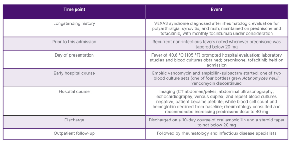
Table 1: Timeline of key clinical events.
VEXAS syndrome: vacuoles, E1 enzyme, X-linked, autoinflammatory, somatic syndrome.
Likewise, the absence of leukocytosis and a procalcitonin of 0.52 ng/mL should not be read as straightforwardly reassuring: tofacitinib pharmacologically suppresses inflammatory markers, so values in this range while receiving JAK inhibition may represent a larger signal than the same values would in an untreated patient. A procalcitonin threshold of 0.5 ng/mL has been reported to suggest bacterial infection among patients with autoimmune disease; however, this figure derives principally from studies in which procalcitonin was specific but only moderately sensitive (approximately 53–67%) for true infection,13 while other studies using substantially lower cut-offs (approximately 0.2 ng/mL and 0.09 ng/mL) were designed to rule infection out rather than in.14,15 This patient’s value of 0.52 ng/mL therefore cannot be treated as unequivocally reassuring, particularly given concurrent JAK inhibition. Recovery of Actinomyces neuii from only one of two culture sets, and negative imaging and repeat cultures obtained after antimicrobial therapy had already begun, are consistent with either contamination/transient bacteremia or an adequately treated true infection, and do not by themselves distinguish the two. On balance, the overall course was judged more consistent with an autoinflammatory flare, but the diagnosis should be regarded as probable rather than definitively established.
The patient’s subsequent course lends indirect support to this interpretation. Over the following 6 months, he was admitted to the hospital several more times with fever in the pattern of his VEXAS flares. Infectious workup was negative and antibiotics were withheld at each of these admissions, yet every episode resolved with glucocorticoids alone. This repeated pattern of culture-negative, antibiotic-free defervescence in the same patient makes it more plausible, in retrospect, that the index admission also represented an autoinflammatory flare with an incidental or transient Actinomyces neuii bacteremia, rather than the first of a series of unrecognized infections. This argument is necessarily retrospective, however, and does not resolve the diagnostic uncertainty that existed at the time of the index admission itself, which remains the focus of this report.
Pancytopenia in VEXAS arises from clonal hematopoietic dysfunction and chronic inflammation. Mutant UBA1 allows a clonal population of hematopoietic stem and progenitor cells to dominate, skewing differentiation toward the myeloid lineage while impairing production of megakaryocytes, erythroid cells, and lymphoid cells.16 This leads to progressive bone marrow failure that worsens independently of the inflammatory flares. Worsening pancytopenia cannot be reliably used as a marker of autoinflammatory flares in VEXAS syndrome; instead, it may reflect clonal evolution, treatment effects, particularly immunosuppression, or disease progression rather than an acute inflammatory flare.
The current treatment of VEXAS remains challenging. JAK inhibitors and IL-6-blocking agents each offer only partial control of symptoms and do not significantly reduce glucocorticoid exposure or prevent hematologic progression.7,17,18 Azacitidine shows promise, with inflammatory response rates of 54% at 12 months and molecular responses in 65% of patients, including those without myelodysplastic syndrome.19 At present, allogeneic hematopoietic stem cell transplantation remains the only treatment that is curative.20,21
LIMITATIONS
Exact clock times for peak fever, blood culture collection, initiation of ampicillin-sulbactam, and escalation of prednisone were not systematically recorded in a way that allows fine-grained sequencing of these events; because the antibiotic and the glucocorticoid change occurred in the same early hospital period, defervescence cannot be attributed with confidence to either intervention alone, or to spontaneous resolution of an autoinflammatory flare.
CONCLUSION
For individuals with VEXAS syndrome, fever may reflect underlying autoinflammation rather than infection. However, improved diagnostic methods are urgently needed to distinguish autoinflammatory flares from infection, as current clinical and laboratory markers, including C-reactive protein and procalcitonin, lack specificity, and consensus guidelines highlight this as a major unmet need. No validated scoring systems currently exist to guide antibiotic use versus immunosuppression in VEXAS; management decisions rely on clinical judgment and the integration of laboratory and imaging findings. Recent reviews have emphasized the importance of developing response criteria and standardized tools to better differentiate autoinflammatory activity from infectious complications in VEXAS. This case highlights the diagnostic complexity where autoinflammatory disease and infection intersect in VEXAS syndrome.