Abstract
Hammer toe is the deformity of a lesser toe characterised by flexion at the proximal interphalangeal joint, starting out flexible but turning rigid with time. In patients who have diabetes, abnormal mechanics of the foot along with an increase in plantar pressures because of this deformity may cause calluses, ulceration, and even skin breakage. This review article will elaborate on the pathophysiology of hammer toe in patients with diabetes along with the techniques of risk assessment of the same problem.
The reviewed literature highlights the significance of chronic hyperglycaemia, advanced glycation end-products production, tendon alterations, neuropathy, weak foot musculature, and abnormal biomechanics in the development of hammer toe. The clinical evaluation comprises inspection of deformity of the toe, skin integrity, plantar pressures, peripheral neuropathy, peripheral vascular disease, and shoes. The risk stratification and adequate documentation will assist in choosing the high-risk patients requiring special care. An early diagnosis, proper footwear, decreased pressures, periodic assessment of the feet, and patient education can prevent the development of ulcers.
Key Points
1. Hammer toe deformity is an established deformity of the feet that develops in people with diabetes. Both neuropathy and biomechanical problems cause weight bearing abnormalities, calluses, and ulcers.2. Early risk assessment and intervention are critical, as identifying neuropathy, altered biomechanics, and footwear-related pressure during the flexible stage can prevent progression to rigid deformity and diabetic foot complications.
3.The use of clinical risk assessments including neuropathy, peripheral artery disease, foot deformity, and previous foot ulcers is important to identify people who have a risk of developing diabetes foot conditions.
INTRODUCTION
Hammer toe is one of the lesser toe deformities that can affect individuals with diabetes. It is a condition whereby the proximal interphalangeal joint of the toes becomes flexed. It is either flexible or rigid, depending on the extent of deformity.1 It often leads to diabetic foot as a result of corns, calluses, and ulcers. If left untreated, it can result in amputations.2 The clinical importance of hammer toe goes beyond the deformity, as it increases the risk of developing foot ulcers, which was documented to be around 6.3% in patients with diabetes overall, and leads to subsequent diabetic foot complications. Furthermore, it was estimated that about 25% of patients with diabetes might develop diabetic foot ulcers in their lifetime, with rates even higher among those patients with structural deformity.3 Hammer toe is one of the many foot deformities that may occur in those with diabetes, highlighting the necessity of conducting a clinical examination of foot problems.
The pathophysiology behind hammer toe is multifaceted, with various neuropathic, biomechanical, and vascular components interacting to result in loss of tissue integrity and progressive deformity.
Neuropathy
In diabetes, diabetic peripheral neuropathy (DPN) leads to loss of protective sensation (LOPS) and motor control, resulting in atrophy of intrinsic foot muscles and motor imbalance due to nerve damage. This muscle imbalance causes the toes to contract abnormally, along with flexion of the proximal interphalangeal joint and extension of the metatarsophalangeal joint.4 Furthermore, glycosylation of muscle tendons causes shortening and stiffening, which exacerbates the foot deformities like hammer toes. Additionally, nerve damage induced by prolonged uncontrolled hyperglycaemia in DPN results in the weakening of intrinsic muscles of the foot, thereby allowing stronger extrinsic muscles to dominate, which further adds to the pathophysiology of characteristic bending in hammer toe.5
Vascular Insufficiency
Peripheral artery disease (PAD) is a major complication in patients suffering from diabetes due to insufficient blood flow to the legs, which results in impaired tissue perfusion and healing. Narrowing of the arteries occurs due to atherosclerosis, resulting in insufficient blood flow to the feet.⁶ PAD cannot cause hammer toe, but it increases the likelihood of skin damage, foot ulcers, infections, and delayed wound healing. This is why these complications, together with structural abnormalities of the feet, make the likelihood of developing diabetic foot problems even higher.
Biomechanical Factors
In diabetes, chronic hyperglycaemia predisposes patients to altered foot structure and gait, which often leads to deformities like hammer toe; changes in foot mechanics in long-standing cases increase pressure in specific areas, causing structural deformities. Hammer toe was found to be associated with elevated plantar pressures and simultaneous muscle atrophy of the intrinsic foot, with major biomechanical changes. Recurrent injury due to diabetic neuropathy leads to these structural changes, where pronation, equinus, or hallux valgus further heightens imbalances, resulting in a less effective windlass mechanism during walking.7 These factors generate a vicious cycle of increased forefoot loading and foot deformity progression.
Hammer toe falls under the category of foot problems that can affect people suffering from diabetes.8 This foot deformity leads to changes in foot biomechanics, resulting in high foot pressure on certain regions of the foot, which subsequently results in skin breakdown and foot ulceration. As a result of high plantar pressures in hammer toe, the efficiency of the windlass mechanism and dorsiflexion is prominently decreased, leading to increased risk of skin ulceration in areas of prominent bone sites.9 Additionally, this results in infections, particularly in cases of impaired wound healing. The major risk factor contributing to infections is the presence of skin ulcers that can pose a significant risk of foot amputation in individuals with diabetes, with hammer toe deformity causing repeated foot trauma and reduced overall mobility. Neuropathy and tissue healing issues could predispose patients to issues of the diabetic foot. Charcot neuro-osteoarthropathy is an issue that comes about due to neuropathy and should not be mistaken as a consequence of hammer toe formation.10
Knowing the high probability of complications due to hammer toe in diabetes, early identification of signs and enforcement of adequate preventive strategies are highly important. The management of hammer toe is preventive: patients wear appropriate footwear with enough space in the toe box, foot screening is performed to identify the deformity, and patients are given education on performing self-exams for issues such as sores or redness.11 This approach to management was found to decrease the onset of foot ulcers and associated complications. Specific interventions such as custom orthotics to correct altered biomechanics and appropriate shoes that reduce peak pressures are notably effective in neuropathic patients with diabetes. Additional interventions include wearing protective socks, controlling blood sugar levels, and trimming nails to prevent injuries.12 Even after the implementation of these recommendations, the existing preventive care for hammer toe remains suboptimal in many healthcare settings, often due to less awareness and restricted access.
The objective of this literature review is to study the pathology of hammer toe in patients with diabetes and examine the risk factors associated with it. This literature review is intended to increase knowledge about the pathophysiology of hammer toe disease.
METHODOLOGY
A narrative literature review was carried out with the objective of reviewing the available evidence concerning the clinical significance, pathophysiology, aetiology, and risk factors associated with hammer toe deformity in patients with diabetes. A comprehensive electronic search was conducted using PubMed, Google Scholar, and Scopus for published articles in the English language up to 2025. Specific search terms were “lesser toe deformities,” “hammer toe,” “diabetic neuropathy,” “diabetes mellitus,” “foot deformity,” “plantar pressure,” “biomechanics,” and “diabetic foot ulcer.” Additionally, reference lists of related articles were also screened manually to detect pertinent studies. Clinical trials, original articles, reviews, observational studies, and guidelines that addressed hammer toe deformity in diabetes were included. Studies targeting particularly non-diabetic populations or lacking clinical plausibility were excluded.
MECHANISM OF HAMMER TOE IN DIABETES
The mechanism underlying the hammer toe deformity due to diabetes is complex, involving molecular, metabolic, and biomechanical changes augmented by chronic hyperglycaemia.13 These alterations result in the functional and structural impairment of ligaments, tendons, and intrinsic foot muscles, which give rise to the disruption of digital alignment eventually.
Role of Advanced Glycation End-Products in Cross-Linking of Collagen
In diabetes, chronic hyperglycaemia causes non-enzymatic glycation of lipids and proteins, resulting in the formation of so-called advanced glycation end-products (AGE).14 The products deposit particularly in the connective tissue of ligaments and tendons. Furthermore, these AGEs create covalent cross-linking of collagen fibres, leading to the decreased elasticity and solubility of collagen, which increases mechanical fragility and tissue stiffness.15 Eventually, the tendons lose their inbuilt ability to adapt to dynamic loading forces, rendering the toes in a constant flexed posture, which is characteristic of hammer toe deformity.
Activation of RAGE-Mediated Oxidative and Inflammatory Pathways
AGEs act through receptors known as receptors of advanced glycation end-products (RAGE), which act as a critical mediator of AGE-associated cellular dysfunction.16 The interaction of AGE–RAGE initiates cascades of downstream intracellular signalling, such as the NF-κB pathway. This leads to the formation of reactive oxygen species and related pro-inflammatory cytokines, augmenting a vicious cycle of oxidative stress and inflammation.17 As a result, cellular damage occurs, resulting in extracellular remodelling, fibrosis, and tendon biomechanical damage.
Mitochondrial Dysfunction in Tendon Cells
The persistent presence of hyperglycaemia results in oxidative stress that leads to malfunctioning mitochondria in the cells of tendons, including tenocytes and tendon stem/progenitor cells.¹⁸ As a result, cellular senescence and cell death take place, resulting in the reduced ability of cell regeneration.¹⁸ In addition, hyperglycaemia results in decreased activity of adenosine monophosphate–activated protein kinase/early growth response 1 (EGR1) signalling pathway that causes low levels of the extracellular matrix proteins such as type 1 collagen, transforming growth factor-beta 1 (TGF-β1) and biglycan.¹⁹ The role of EGR1 is extremely important for tendon differentiation and repair.²⁰
Intrinsic Muscle Damage and Neuromuscular Equilibrium Disruption
Diabetic neuropathy complicates the deformity, as it results in intrinsic foot muscle atrophy caused by denervation that helps to stabilise the toes normally.21 Further, it has been noted that the dysfunction of muscles in the feet due to ageing is linked with the dysfunction of muscles in diabetic feet.22 This leads to intrinsic foot muscle weakening, resulting in unopposed extrinsic foot muscle action that pulls the toes into a flexed position.23 This disruption in balance further elevates joint misalignment, resulting in fixed deformity over time.
Integrative Pathophysiology
Together, these molecular and cellular mechanisms create a pathophysiological sequence, where RAGE-mediated chronic inflammation, AGE accumulation, oxidative stress-induced mitochondrial damage, and disrupted neuromuscular balance interact synergistically.24 The resultant loss of tendon elasticity, disrupted muscle coordination, and poor structural integrity damage normal foot mechanics, leading to the gradual onset and progression of hammer toe deformity in diabetes.25
The mechanism (Figure 1) underlying the hammer toe in diabetes is not only structural, but also due to chronic metabolic dysregulation. Hence, hammer toe in diabetes appears to be due to the intersection of biochemical, cellular, and mechanical pathways, denoting the complex intersection of metabolic control and musculoskeletal health.26
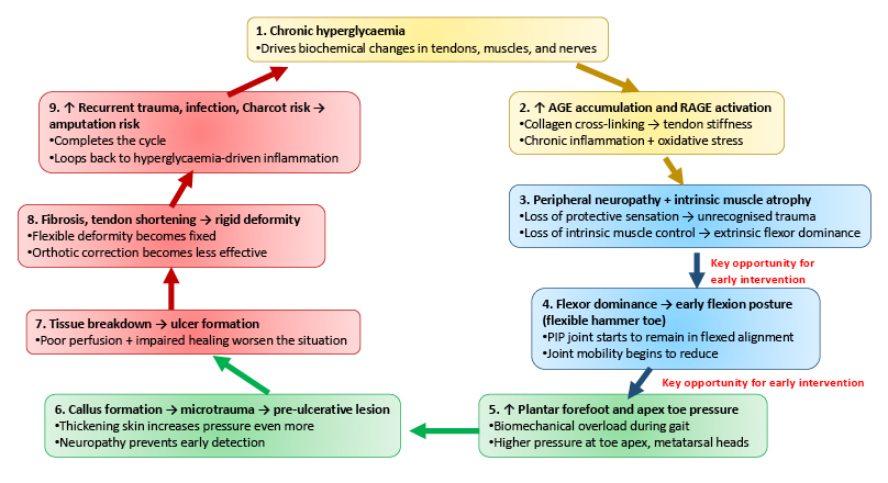
Figure 1: Vicious cycle of hammer toe progression in diabetes.
A loop of hammer toe progression in which chronic hyperglycaemia promotes the accumulation of AGE- and RAGE-driven inflammation, causing neuropathy, intrinsic muscle atrophy, flexor dominance, and tendon stiffening. These alterations increase plantar pressure at the toe apex, promoting callus formation, microtrauma, and subsequently ulceration. Tendon shortening and progressive fibrosis turn flexible deformity into rigid hammer toe, increasing the risk of repeated ulceration, infection, and amputation. Disrupting this vicious cycle via proactive intervention is required to prevent irreversible deformity and limb threatening complications.14-26
AGE: advanced glycation end-products; RAGE: receptors of advanced glycation end-products; PIP: proximal interphalangeal.
RISK ASSESSMENT
Rationale Behind Risk Assessment
Hammer toe results in changes in normal plantar pressure distribution, leading to the formation of calluses and subsequently skin breakdown. Studies have highlighted the persistent relation between diabetic foot deformities and the formation of foot ulceration and high risk of amputation. The critical components of comprehensive diabetic foot care consist of early identification of signs related to hammer toe and risk assessment, aiming to halt diabetic ulcer formation and related morbidity.27
Risk Assessment Objectives
The primary objectives in hammer toe risk assessment are to identify high-risk individuals, to categorise them according to risk factors, and to improve specific proactive interventions.28 Risk assessment consists of detecting modifiable extrinsic risk factors, such as areas in the foot with high plantar pressure or improperly fitting footwear. Additionally, it involves identification of intrinsic factors such as diabetic neuropathy, PAD, and loss of muscle balance.29 Integrating these examinations helps to identify and improve preventive measures, optimise follow-up frequency, and decide when referral to orthopaedic or podiatry evaluation is required.
Important Risk Factors
In hammer toe, there are multiple risk factors with both intrinsic and extrinsic components.30 Intrinsic components include diabetic neuropathy that leads to LOPS and recurrent unrecognised injury and peripheral artery disease, causing impaired tissue perfusion and poor wound healing.31
Additionally, structural changes like restricted joint mobility, hammer toes, clawed toes, and wasting of intrinsic muscles lead to abnormal foot pressure distribution.32 Extrinsic components like elevated focal plantar pressures and poor-fitting footwear could drive foot deformity and skin breakdown.33 Furthermore, a history of foot ulceration or amputation persists as a strong future risk of recurrent foot ulcers. These components collectively interact through vascular, biomechanical, and metabolic pathways to result in progressive foot deformity and ulceration. 34,35
Comprehensive Clinical Risk Assessment
Clinical risk assessment begins with a focused history including symptoms due to neuropathy, footwear habits, previous ulcers, and deformity progression.36 Physical examination comprises structured examination of calluses, erythema, digital deformity, and skin breakdown.37 For instance, the identification of calluses under metatarsal heads or toe tips is highly important, as it shows localised pressure points and a high risk of ulcers. Additionally, a comprehensive neurological examination including 10 g monofilament test, vibration test, and temperature test helps to detect the loss of protective sensation.38 Vascular assessment includes ankle-brachial index (ABI) measurement, palpation of all peripheral pulses, and toe pressure assessment to diagnose PAD.39 Biomechanical examination consists of the assessment of toe flexibility and gait assessment to identify functional disability and severity of deformity.40
Objective Investigations
If the findings from comprehensive assessment show pressure overload or prominent deformity, objective investigations are highly recommended. Assessment of plantar pressure offers quantitative data on focal stress points, which helps in planning offloading interventions.41 Furthermore, imaging like weight-bearing radiographs provides complete information on bony alignment and joint subluxation, while ultrasonography or MRI could be useful in detecting tendon pathology or soft tissue complications.42 In persons with a suspected complication, non-invasive vascular investigations such as toe pressure measurement or duplex ultrasonography are helpful to aid further management.43
Risk Stratification
According to the International Working Group on the Diabetic Foot (IWGDF) guidelines 2023, people with diabetes are grouped into four classes depending on the risk of foot ulcers. People with no LOPS or PAD belong to category 0, which is very low risk. People with LOPS or PAD are grouped into low risk and belong to category 1. Foot deformity alone, including hammer toe, in the absence of LOPS and PAD remains in category 0, whereas PAD alone is classified as category 1.44 People who have LOPS and PAD, LOPS and foot deformities, or PAD and foot deformities are grouped into moderate risk and belong to category 2. Lastly, people with LOPS or PAD and previous foot ulcers, amputations of the lower limbs, or end- stage renal diseases are grouped into high risk and belong to category 3.44 Foot screenings are recommended every year for category 0, every 6–12 months for category 1, every 3–6 months for category 2, and every 1–3 months for category 3.44
Documentation of Risk Assessment
Since hammer toe deformity in diabetes is an important yet modifiable risk factor for foot ulceration, it is key to properly document risk assessment to improve patient care, which helps to make informed management choices.45 This systematic approach of maintaining a record of deformity type (rigid or flexible), callus assessment findings, neuropathy findings, vascular assessment findings, and evaluation of footwear during each visit contributes to an enhanced continuity of care.46
Depending upon the risk category, re-assessment intervals must be planned. This structured assessment framework consisting of biomechanical, neurological, and vascular evaluation and follow-up might decrease the incidence of ulcers and help in risk stratification and early detection of diabetic foot complications.
CONCLUSION
Hammer toe is a condition which can be classified as a musculoskeletal condition in patients with diabetes. It results from the complex interaction of chronic uncontrolled blood glucose, diabetic neuropathy, impaired perfusion, and disruption of biomechanical balance. As a result, it leads to abnormal distribution of plantar pressure, thereby heightening the chances of callus formation, skin breakdown, infection, and limb-threatening complications such as amputation risk. This literature review emphasises the importance of understanding the pathophysiology of hammer toe deformity as well as identifying those risk factors associated with the development of the deformity. Assessment of the risk factors through neurologic, vascular, and biomechanical examinations will assist in determining which patients are at risk for diabetic foot disorders.