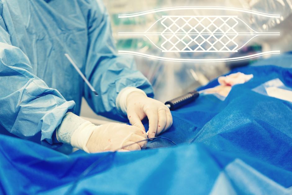
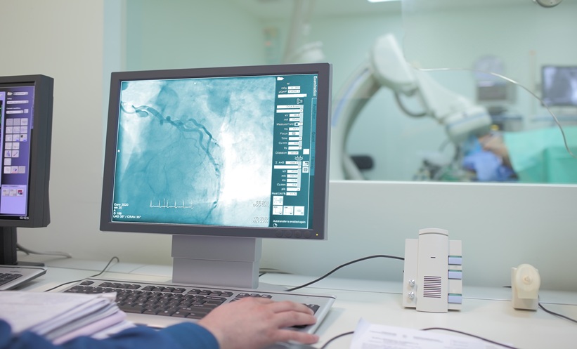
MRI-GUIDED stent implantation for aortic coarctation demonstrated technical and procedural feasibility in a multistage preclinical study, with successful implantation achieved in all five animal models.
The findings highlighted the potential for magnetic resonance imaging (MRI) to support radiation-free endovascular treatment of aortic coarctation, although further investigation is required before clinical implementation.
MRI-Guided Stenting Evaluated in Preclinical Models
Researchers assessed the feasibility of MRI-guided stent implantation for aortic coarctation using a structured three-stage preclinical evaluation protocol.
The study investigated a commercially available MR Conditional guidewire alongside dedicated research prototypes, including nitinol stents, delivery systems, and balloon catheters.
Initially, device visibility was assessed using a standardised water phantom at magnetic field strengths of 1.5 T and 3.0 T.
Researchers subsequently evaluated procedural performance in a polymer-based aortic coarctation vessel phantom, where 10 stent implantations were performed by six novices and four experts.
Four additional balloon dilatations were performed by experts to assess procedural feasibility.
The final evaluation involved MRI-guided stent implantation in five porcine models, providing an opportunity to assess the technique under preclinical in vivo conditions.
Successful Stent Implantation Across Animal Models
All devices were visible under MRI at both magnetic field strengths, although the extent of imaging artefacts varied between components.
Lateral artefact widths ranged from 5.3–7.8 mm for the guidewire, 20.9–38.5 mm for the stent delivery system, 13.1–19.9 mm for expanded stents, and 17.4–30.5 mm for the balloon catheter.
During phantom testing, mean stent implantation duration was 10.2±7.2 minutes for novices, compared with 6.2±2.7 minutes for experts.
MRI-guided stent implantation was subsequently successful in all five animal models, with a mean procedure duration of 13.6±7.5 minutes.
In one animal, researchers additionally performed balloon post-dilatation, demonstrating the procedural feasibility of this supplementary intervention.
Potential for Radiation-Free Aortic Coarctation Treatment
The successful completion of MRI-guided stent implantation across the preclinical evaluation stages supported the technical feasibility of this approach.
Device visibility at both tested magnetic field strengths, alongside successful implantation in animal models, provided preliminary evidence supporting further development.
However, the findings were limited to phantom testing and animal experiments, meaning clinical performance in patients remains unestablished.
Overall, the researchers concluded that MRI-guided stenting represents a feasible preclinical approach to aortic coarctation treatment, supporting further investigation towards clinical translation of radiation-free endovascular interventions.
Reference
Bajestani DG et al. Multistage Preclinical Evaluation of MRI-Guided Stenting for Coarctation of the Aorta using in vitro and Porcine Models. Cardiovascular and Interventional Radiology. 2026.