CONTRAST-enhanced mammography showed good accuracy for assessing breast cancer response after neoadjuvant therapy in pooled data.
A systematic review and meta-analysis of 15 studies involving 793 patients found that contrast-enhanced mammography may offer a clinically useful option for evaluating pathological complete response after neoadjuvant therapy, with diagnostic performance described as comparable to MRI.
Neoadjuvant therapy is increasingly used in breast cancer management to downstage tumors, support breast-conserving surgery, and provide an early measure of treatment response. While MRI remains the reference standard for response assessment, cost, access, acquisition time, and patient tolerance can limit its use. Contrast-enhanced mammography combines mammographic anatomical detail with contrast-based functional imaging, enabling visualization of tumor enhancement in a single examination.
Diagnostic Accuracy Supports Clinical Potential
Using surgical histopathology as the reference standard, pooled analysis showed a diagnostic odds ratio of 9.30, with substantial heterogeneity across studies. Sensitivity was 74%, while specificity was 82%, indicating that contrast-enhanced mammography had moderate-to-high performance for identifying pathological complete response after neoadjuvant therapy.
The summary receiver operating characteristic curve showed an area under the curve of 0.83, supporting good overall diagnostic accuracy. Based on a reported pathological complete response prevalence of 31.7%, positive predictive value was 66% and negative predictive value was 87%. This high negative predictive value suggests that absence of abnormal enhancement on contrast-enhanced mammography may help reduce the likelihood of residual disease.
Findings Compared With MRI
The analysis reinforces contrast-enhanced mammography as a practical imaging approach when MRI is unavailable, contraindicated, poorly tolerated, or less accessible. Its ability to provide both anatomical and functional information may be particularly valuable after neoadjuvant therapy, when clinicians need to distinguish residual disease from treatment-related changes.
However, the findings also highlight limits. The moderate positive predictive value means contrast-enhanced mammography should not be used alone to confirm pathological complete response. Histopathologic assessment remains essential, particularly because residual nodal disease and subtle tumor bed changes may not be fully captured by imaging.
Heterogeneity was initially substantial, although removal of one study increased the diagnostic odds ratio to 10.28 and reduced heterogeneity to 19.4%. Differences in tumor subtype distribution, neoadjuvant therapy regimens, imaging protocols, and pathological complete response definitions may have influenced pooled estimates.
Further prospective, multicenter studies are needed to standardize acquisition protocols, interpretation criteria, and histopathologic definitions. For now, contrast-enhanced mammography appears to be a promising, accessible tool for post-neoadjuvant therapy breast cancer response assessment, particularly as part of a multimodal strategy rather than a standalone substitute for pathology.
Reference
Galati F et al. Diagnostic accuracy of contrast-enhanced mammography in evaluating breast cancer response to neoadjuvant therapy: a systematic review and meta-analysis. Radiol Med. 2026. doi:10.1007/s11547-026-02215-y.
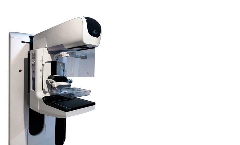
Featured Image: samunella on Adobe Stock.
- Author: