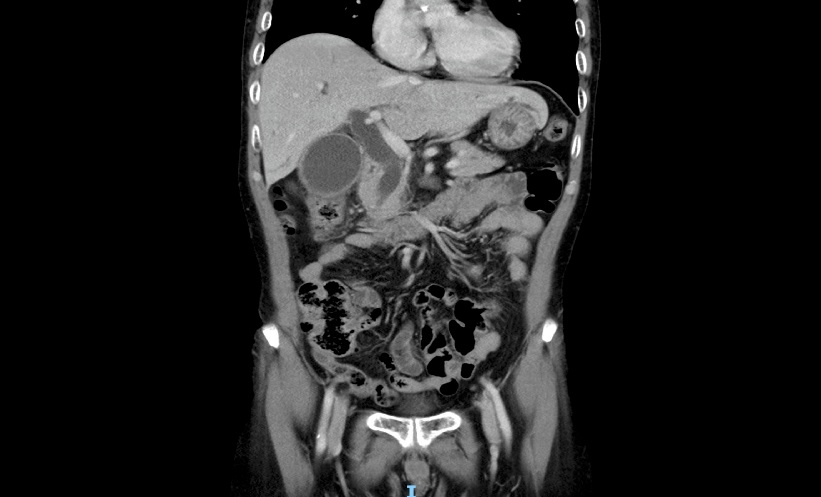
CT HISTOGRAM analysis could provide complementary information for assessing liver fibrosis in patients with chronic hepatitis B, according to a study comparing non-contrast CT findings with biopsy results, FibroScan measurements and serum-based fibrosis scores.
While FibroScan showed the strongest relationship with histological fibrosis, several CT histogram parameters also correlated with established measures of disease severity.
Liver fibrosis, a build-up of scar tissue caused by ongoing hepatic injury, is commonly assessed using liver biopsy alongside non-invasive techniques. The findings suggested quantitative information derived from routine non-contrast CT scans could complement these established assessment methods without replacing them.
CT Histogram Analysis Showed Links with Fibrosis Measures
The study included 40 patients with chronic hepatitis B who had histopathologically staged liver fibrosis. Histogram parameters derived from non-contrast CT images were compared with biopsy-based Ishak fibrosis stage, FibroScan measurements and the serum-based APRI and FIB-4 scoring systems.
Among the histogram features evaluated, Size%L, standard deviation and variance demonstrated significant positive correlations with the Ishak fibrosis score. Size%L also correlated positively with APRI and FIB-4, alongside biopsy-based fibrosis staging. However, FibroScan demonstrated the strongest relationship with histopathological fibrosis staging across all methods assessed.
Potential Role Alongside Established Assessment Tools
The findings indicated that CT histogram analysis reflected structural characteristics of liver fibrosis and showed significant correlations with both biopsy findings and established non-invasive assessment methods.
Rather than replacing liver biopsy or elastography, the technique may provide complementary quantitative information during routine imaging. Because the analysis used non-contrast CT images, it may offer additional value in patients already undergoing abdominal CT for other clinical indications.
Adjunctive Imaging Marker Warrants Further Evaluation
The researchers concluded that CT histogram analysis may have potential as an adjunctive imaging marker in chronic hepatitis B. Although the technique demonstrated significant associations with fibrosis staging and serum-based scores, FibroScan remained the method most closely aligned with histopathological findings.
The findings support further evaluation of CT histogram analysis as an adjunctive imaging marker that could add quantitative fibrosis information to routine abdominal CT examinations.
Reference
İspiroğlu M et al. Comparative evaluation of non-contrast CT histogram analysis, FibroScan, and serum markers for liver fibrosis assessment in chronic hepatitis B patients. Clin Radiol. 2026;DOI:10.1016/j.crad.2026.107425.
Featured image: Issara on Adobe Stock