Walter Maksymowych| Spondyloarthritis Research Consortium of Canada, Edmonton; Division of Rheumatology, Department of Medicine, University of Alberta, Edmonton; CARE Arthritis Ltd, Edmonton, Alberta, Canada
Citation: AMJ Rheumatol. 2026; https://doi.org/10.33590/rheumatolamj/R4A0H99M

Your investigative career is characterized by an interdisciplinary partnership with musculoskeletal radiologist Robert Lambert. How did that collaboration develop, and how did working closely with a radiologist alter your diagnostic reasoning at bedside?
When I started doing research in axial spondyloarthritis (axSpA), I realized that I needed an objective way of documenting the severity of disease. I needed an objective way to document the extent of inflammation, its severity, and also the extent and severity of joint damage.
It was around that time, when I began my career in research, that data was starting to appear, and I realized that MRI would be very helpful in interpreting and identifying disease activity in spondyloarthritis, specifically in the sacroiliac joints and the spine. Then, I thought that it would be very helpful to engage with a radiologist who might be interested in collaborating with me, teaching me about MRI and how it might be useful in evaluating spondyloarthritis. That’s how I met Robert Lambert. I cannot now imagine working in the clinic and evaluating a patient’s back pain without having access to medical images. It’d be like working while blindfolded.
axSpA is characterized by a balance between localized bone erosion and paradoxical osteoblast proliferation leading to ankylosis. What do you consider the most promising cellular target for arresting syndesmophyte bridging without compromising systemic bone mineral density?
The whole process begins with inflammation. No matter what the stimulus that triggers the inflammation is, it’s crucial that we intervene and alleviate the inflammatory process. Without alleviation of inflammation, it’s going to be very difficult to arrest development of syndesmophytes and bridging bone. We have growing evidence for this from observational cohort studies. It’s very difficult to examine this in a randomized, placebo-controlled setting, because trials take a long period of time and you can’t keep patients on placebo for an extended period of time. Additionally, we do have growing evidence that there is a link between inflammation and new bone formation. What is interesting is that, for the longest time, we thought that T cells might not be important; that other cells, for example, cells of the innate immune system and macrophages, may be more important. To me, it seems like macrophages are still important, because they have a lot of the biochemical machinery that promotes inflammation. Attention in the field has recently turned back to T cells. There is growing evidence that T cells are recognizing antigens, specifically self-antigens, and that self-antigen is being presented by B27 molecules on antigen presenting cells. There is a process of cross-reactive autoimmunity where external antigens that resemble self-antigens, for example, those derived from bacteria, are triggering the activation of a very restricted set of T cells. We’ve seen evidence for these clonotypes in several tissues of patients with this condition. It’s quite possible that if we can intervene, for example, with monoclonal antibodies and block this specific group of T cells that are being activated, even though they may be only a small portion of the T cell repertoire, that we might be able to intervene in the process. So, T cells are back! The other cells may still be important, but the field is starting to realize that we should be targeting T cells.
Deep learning systems are being trained to automatically detect sacroiliitis and spinal syndromophocytes on MRI and CT. Do you anticipate AI evolving into an autonomous diagnostic modality in clinical practice, or will anatomical variability keep it restricted to research quantification?
I think that AI will increasingly play a very important role in the interpretation of imaging in an adjunctive way. AI will, at the very least, be able to identify if there is an abnormality, so there will be a more restricted number of images that will be directed to radiologists. This is helpful, as it helps radiologists avoid spending a lot of their time looking at normal scans. Now, there are a lot of findings on MRI of the sacroiliac joints and the spine that may not necessarily be connected with spondyloarthritis. Sometimes, these abnormalities can be difficult to distinguish from what we see in spondyloarthritis. That’s the hurdle, and why I think it will still be some time before AI can replace the radiologist. Eventually, if we have enough ground truth and there’s enough training of neural networks, we may get to a point where AI gets pretty good. So, who knows? Technology is accelerating very rapidly, but I don’t think we’re close to it yet. However, I do think it has huge potential for the future.
The screening for the SASPIC cohort revealed significant variations in diagnostic categorization after systemic MRI evaluation. How frequently did systemic imaging reverse initial clinical diagnoses in patients presenting with undiagnosed back pain?
That’s a very important question, because patients present with back pain to a variety of specialists. When they present with iritis, inflammatory bowel disease, or psoriasis, those patients should be referred to a rheumatologist, especially if they are B27-positive.
In this graph we can see what the impact of MRI really is (Figure 1). It displays the frequency of cases that are diagnosed with spondyloarthritis. You can see it’s very close to 100% in patients with psoriasis, uveitis, and inflammatory bowel disease after clinical assessment. Then, they get the results of the labs and the radiography and the frequency of axSpA goes down. The green bars show the frequency of axSpA when rheumatologists then get the results of the MRI assessment. You can see that, especially for the B27 negative cases, the proportion of cases diagnosed with axSpA has dropped off. Essentially, MRI is helping to rule out axSpA. Without MRI, we would be over-treating a large number of patients.
One further really important point I will make is there are no clinical features that help to distinguish back pain that is associated with axSpA versus back pain that is non-axSpA. The only discriminatory features are B27 and imaging. MRI has a profound impact on the diagnostic accuracy. In other words, you can’t rely on other clinical features like the presence of peripheral arthritis, enthesitis, psoriasis, or uveitis when patients are presenting with back pain. The only discriminatory features are B27 and MRI imaging.
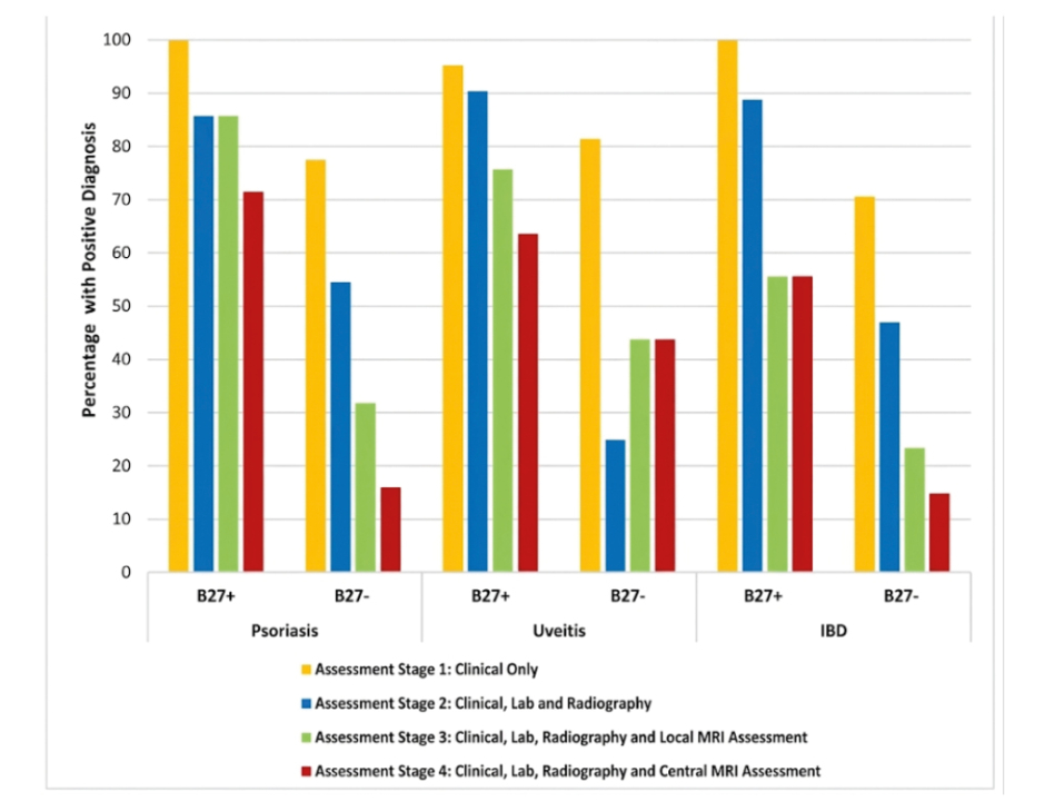
Figure 1: Percentage of patients from SASPIC 2 diagnosed with axSpA across referral sources, HLA B27 status, and sequential assessment stages (N=151).1
Screening for axSpA in patients presenting with PsO, AA, or IBD and undiagnosed chronic back pain. HLA-B27 is positive in only 40–50% of axSpA cases presenting with PsO or IBD, and no clinical features help distinguish between a diagnosis of axSpA and non-axSpA. Bars represent the proportion of positive diagnoses confirmed across Sequential Assessment Stages 1–4. Because MRI has a profound impact on diagnostic accuracy, it can be concluded that all patients should have MRI SIJ evaluation.
Adapted from Maksymowych et al.1
AA: acute anterior uveitis; axSpA: axial spondyloarthritis; HLA-B27: human leukocyte antigen B27; IBD: inflammatory bowel disease; PsO: psoriasis; SIJ: sacroiliac joint.
Through CARE Arthritis, Edmonton, Canada, you expanded your educational reach to a global community of rheumatologists and radiologists. What remains the most persistent barrier to standardizing imaging interpretation in community practice, and how can digital platforms address it?
I’m going to approach the answer from the perspective of the radiologist and then from the perspective of the rheumatologist. First of all, radiologist training curriculums, whether undergraduate, postgraduate, or fellowship training, do not cover very much spondyloarthritis, if any at all. So, what we can provide through CARE Arthritis is a very large library of case-based content where there is medical imaging with examples of both straightforward and more difficult cases. There are also examples of mimics of spondyloarthritis. All these MRI and X-ray images are available as case-based content on the CARE Arthritis website.
What is also very important is that we provide open-access information to radiologists as to the optimal approach to the imaging evaluation. In other words, how the MRI should be done to optimally evaluate the sacroiliac joints and spine. That information is based on an international consensus of musculoskeletal radiologists from around the world. This includes radiologists who are members of the Society of Skeletal Radiologists (SSR), Spondyloarthritis Research and Treatment Network (SPARTAN), and also the Assessment in SpondyloArthritis International Society (ASAS).
The rheumatologist perspective is that rheumatologists really need to understand the basic language of MRI if they’re going to have any hope at all of interpreting radiologists’ reports, in which radiologists use a lot of technical language. If rheumatologists are not familiar with lesions and the diagnostic capacity of the different lesions that are observed on MRI, it’s going to be very difficult for them to apply the information for diagnostic interpretation. Therefore, it’s very important that rheumatologists learn to understand MRI. Over the years, rheumatologists have learned how to interpret radiography, so all rheumatologists are going to be looking at radiographs of the sacroiliac joint. They should apply the same process to MRI interpretation, get used to looking at MRI images, and be used to talking to radiologists about MRI images. It starts with an understanding of the basic language.
When you co-founded the Spondyloarthritis Research Consortium of Canada (SPARCC), multicenter rheumatology research in Canada was largely fragmented across independent provincial divisions. What structural hurdles did you face in establishing a shared research protocol and standardized imaging repository across Canadian academic sites?
I’ll first tackle the issue of a shared research protocol. We did not have too many difficulties getting this established. The main one was the issue of feasibility. We’re all enthusiastic researchers and want to capture as much data as possible, but a lot of the data, and particularly the standardized questionnaires of patients, are self-reported. That means it takes up time from the patient, but we were able to overcome that by being reasonable with our requests. First, we worked to ensure that patients weren’t spending more than an hour completing their questionnaires. Secondly, in organizing patient visits to the clinic, we would ask for patients to come prior to their appointment and complete their questionnaires. The results of those questionnaires were very helpful at the time to rheumatologists in patient care. It was a win-win situation for patients and for the rheumatologists who are doing the research and providing care at the same time.
Now, with respect to imaging standardization, that was more of a challenge, because imaging is frankly done in a highly variable manner. It doesn’t matter whether you consider community-based sites, academic sites, or anything else; everybody thinks they’re doing the best possible imaging. That required a lot of conversations with radiologists, along with intense effort to persuade radiology teams that this was actually in their best interest. Most importantly, that it was in the best interest of patients, because the images that we requested were those that provided us with the best opportunity to evaluate the sacroiliac joints and spine. Spine imaging is fairly straightforward, but what really needed to be done more appropriately was the evaluation of the sacroiliac joints.
Despite widespread access to MRI and targeted biologics over the past 20 years, epidemiological registries still document an average diagnostic delay of 7–8 years in axSpA. What structural barriers within primary care referral pathways and medical training continue to perpetuate this delay?
There are some reasons for the delay that have existed for decades, which continue to exist and have not been addressed. It begins at medical school and the limited training that students get in rheumatology as a whole, but, specifically, in the interpretation of back symptomatology. It’s hit-and-miss to what degree residents will get exposed to patients with spondyloarthritis. Then, we have a very large number of healthcare practitioners who see patients with back pain. Many of these healthcare practitioners have limited expertise in rheumatological conditions. Most importantly, it pertains to primary care physicians. There’s limited understanding of what constitutes inflammatory back pain, so patients often end up cycling between primary care physicians, physiotherapists, orthopedists, chiropractors, naturopaths, and so on. It’s almost a public health message. It should be standard that, if there is a young person with the onset of symptoms below the age of 45 years with chronic back pain, especially if there’s any nocturnal awakening, it should always be a red flag and these patients should be referred to a rheumatologist. Additionally, there is a role for B27 screening in such patients. Of course, it shouldn’t be done without carefully selecting which patients should be screened, but a message I have to primary care practitioners is, that if you have a young patient in front of you with nocturnal back pain, especially with prominent symptoms in the morning, get a B27 done. If they’re positive, refer to a rheumatologist. This would help reduce this diagnostic delay. Finally, I want to point out that it does occur in women, and the diagnostic delay is even worse. We shouldn’t forget that this disease does occur frequently in women, and the type of back pain I have described in a young woman should result in prompt referral to a rheumatologist. It’s a fact that the disease does occur more frequently in men, but it’s not widely appreciated that the ratio of men to women is not 3:1 or 4;1 as is commonly thought, but it’s probably closer to 1.5:1. Not a very large difference, but interpretation of symptomatology in women is very difficult, which is why young women with chronic back pain should be sent to a rheumatologist.
The SASPIC study showed that patients often enter the healthcare systemvia an ophthalmologist, gastroenterologist, or dermatologist for associated manifestations. What interdisciplinary triage protocols are most effective for identifying spinal disease early among patients present with recurrent uveitis or inflammatory bowel disease?
It’s a great question, as there’s a relationship aspect to it. In other words, I think rheumatologists should cultivate relationships with other health care practitioners and facilitate referral pathways. I’ve worked with dermatologists, gastroenterologists, and ophthalmologists, and they know that I am not going to have patients sitting around for 9 months on the waiting list to see me if they present with these healthcare conditions and with chronic back pain; I’m going to see these patients sooner. What may expedite that referral is if the patient is B27-positive, especially if they have low back and/or buttock pain. There’s no reason why the patient should not have a pelvic radiograph. If the patient is B27-positive and already has some abnormalities on the pelvic radiograph, I’m going to see the patient much sooner, but even if they don’t, I’m still going to triage them earlier than I would a patient referred from the general community with back pain.
What do you hope will be the lasting legacy of your work for patients living with axSpA?
When I reflect on that, there’s a couple of things that stand out. The first is that I have been heavily involved with imaging for early diagnosis, so I hope that through my work with imaging, patients will get an earlier diagnosis. I’ve also been lead investigator for the development of recent classification criteria for this disease, and I hope that patients will be more accurately identified. For 30 years, I have participated in clinical trials as both an expert advisor and a clinical investigator aimed at helping to develop new therapeutics for patients. I hope that this will lead to earlier identification of patients and a better array of new therapeutic options with much greater effectiveness for this condition than we have had in the past. Finally, and this belongs to the theme of earlier diagnosis, I have worked on a new blood test regarding antibodies to 14-3-3 protein η (1443 ETA), which show very promising potential as diagnostic biomarkers for spondyloarthritis. At the end of my career, I hope to be able to say that I’ve done something to help diagnose patients much earlier in their disease course and to facilitate more accurate identification of such patients so that the work that I’ve contributed to in clinical trials can be relevant to the development of new therapeutics.