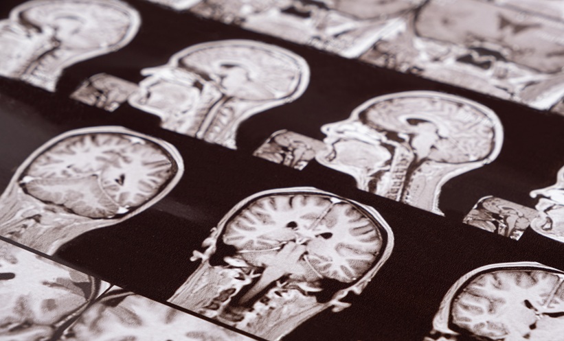
PEOPLE with severe asthma showed differences in brain structure compared with people whose asthma was well controlled. Brain MRI findings from the CONTENTed study suggest that severe type 2 eosinophilic asthma may be associated with changes beyond the airways, although the cause and clinical significance of those changes remain uncertain.
The analysis compared 50 patients with biologic-naïve, type 2-high severe eosinophilic asthma with 25 patients who had mild or moderate, well-controlled asthma. All 75 participants underwent multimodal brain MRI. The scans included quasi-diffusion imaging, a technique used to assess the microscopic structure of white matter, which contains pathways connecting regions of the brain.
MRI Findings in Severe Asthma
Compared with the well-controlled group, patients with severe asthma had lower global white matter volume and lower volumes in the left temporal lobe, thalamus, and globus pallidus. These group differences were statistically significant at p<0.05 after adjustment for age and sex.
Measures of white matter microstructure also differed. The severe asthma group had lower fractional anisotropy and higher mean-alpha values, findings the investigators described as disrupted white matter structural integrity. A separate analysis showed that the differences were widespread across white matter, rather than confined to one small area.
As expected, the groups also differed significantly in fractional exhaled nitric oxide (FeNO), blood eosinophil counts, and exacerbation frequency. Those clinical differences help define the groups being compared, but they do not establish why their MRI findings differed.
What the Findings Mean for Care
Within the severe asthma group, the relationship between FeNO and the white matter imaging measures was a nonstatistically significant trend. The study therefore does not demonstrate that higher type 2 inflammation causes changes in brain structure. Nor did this baseline imaging analysis establish that the MRI differences affect memory, thinking, or other neurological outcomes.
For clinicians, the findings raise a question about the wider effects of severe eosinophilic asthma. They do not support using brain MRI to assess asthma or changing treatment on the basis of these scans. Researchers are following participants to examine whether the imaging findings change after targeted treatment, which may help clarify their significance.
References
Nava GW et al. Structural brain imaging in biologic naïve T2-high severe asthma, baseline data from the CONTENTed study. Abstract 59618. European Respiratory Society Congress, September 5–9, 2026.
Respiratory Therapy. Severe asthma linked to measurable differences in brain structure. September 9, 2026. Available at: https://respiratory-therapy.com/disorders-diseases/chronic-pulmonary-disorders/asthma/severe-asthma-linked-brain-structure/. Last accessed: September 24, 2026.
Featured Image: Zarina Lukash on Adobe Stock.