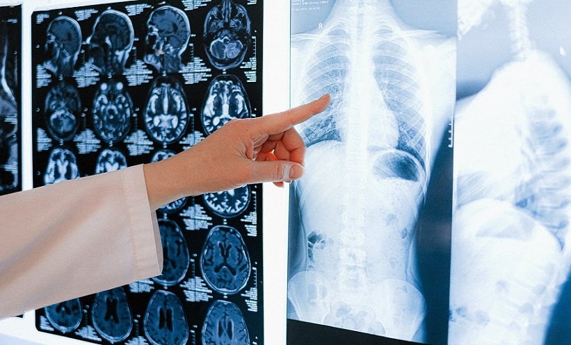
COMPUTER-AIDED detection chest X-ray demonstrated diagnostic performance comparable to experienced radiologists for pulmonary tuberculosis in low-resource, high-burden settings, according to a retrospective pilot study of nearly 500 patients.
Study Context and Rationale
Tuberculosis remains a major global health challenge, particularly in settings where access to radiologists is limited. Computer-aided detection chest X-ray tools have been proposed as a way to support physicians in interpreting chest X-rays for pulmonary tuberculosis. The present study evaluated whether such software could aid clinical decision making where radiological expertise is scarce.
Researchers conducted a retrospective analysis of chest X-ray films collected between 1 January 2017–30 March 2018 in Guinea-Bissau and Ethiopia. Images were assessed by computer-aided detection chest X-ray software and independently reviewed by two experienced Ethiopian radiologists. To enhance relevance for low-resource environments, the analysis also included images captured using mobile phones and a digital camera. Final pulmonary tuberculosis diagnosis based on clinical or laboratory findings, including Mycobacterium tuberculosis detection using Xpert MTB/RIF, served as the reference standard.
Diagnostic Performance of Computer-Aided Detection Chest X-Ray
A total of 498 chest X-rays from patients presenting with symptoms suggestive of tuberculosis were included. Radiologist A identified 50 images as indicative of pulmonary tuberculosis, radiologist B identified 99, and the computer-aided detection chest X-ray software identified 81.
For Xpert-confirmed pulmonary tuberculosis, the area under the receiver operating characteristic curve for the software was 0.84. At a predefined cut-off of 0.5, sensitivity and specificity were: 76.5%; 85.9%, respectively. Radiologist A showed sensitivity and specificity of: 64.7%; 91.9%, while radiologist B demonstrated: 76.5%; 82.3%.
Agreement Between Readers and Software
Agreement with regard to tuberculosis-related findings was moderate. Combined agreement between the two radiologists was κ=0.45, while agreement between each radiologist and the computer-aided detection chest X-ray software was κ=0.56.
The findings indicate that computer-aided detection chest X-ray performs comparably to experienced radiologists, even when applied to images photographed from chest X-ray films using commonly available devices. This suggests potential value for supporting tuberculosis diagnosis in resource-constrained settings where radiology services are limited.