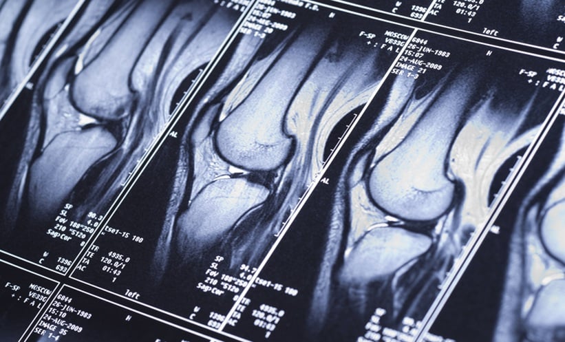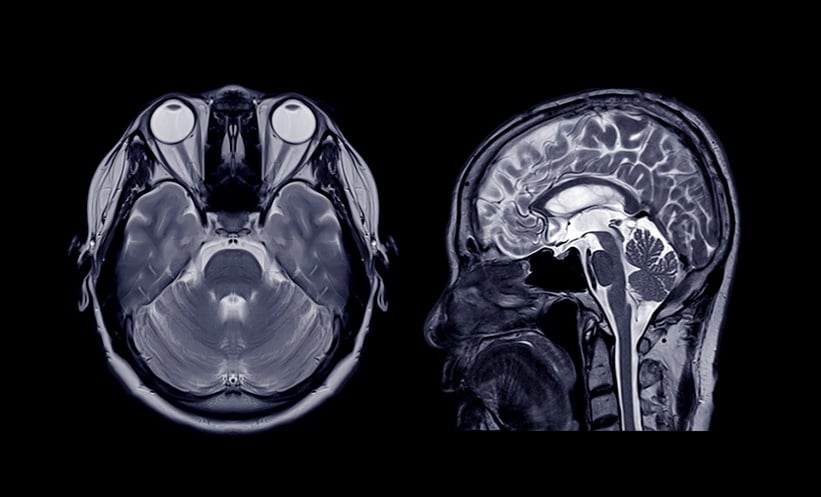
A NEW AI foundation model for neuroimaging was able to read and diagnose brain MRI scans in seconds, a 2026 health system-wide study has found.
Prima, developed by researchers at the University of Michigan, outperformed state-of-the-art general and medical AI models.
AI Model Development
Researchers proposed that the global demand for MRI studies has steadily increased, straining health systems, prolonging turnaround times, and intensifying physician burnout. Such challenges disproportionately affect patients in low-resource and rural settings.
Using data from a large academic health system, researchers developed Prima to support real-life, clinical MRI studies.
AI Model Testing Outcomes
The AI model was trained on more than 220,000 MRI studies and 5.6 million imaging sequences. It was then tested in a 1-year health system-wide study that included nearly 29,500 MRI studies.
Prima outperformed other advanced AI technologies, achieving a mean diagnostic area under the curve of 92% across 52 radiologic diagnoses from major neurologic disorders, including strokes, brain tumours, and haemorrhages. The model was able to identify neurologic conditions with an accuracy of up to 97.5%.
In addition to identifying disease, it was able to offer explainable differential diagnoses, worklist priority for radiologists, and clinical referral recommendations. The system was designed to notify the most appropriate subspecialist.
Prima also incorporated patients’ clinical histories and the reasons why the relevant healthcare professional ordered each MRI. Feedback was immediately available post-imaging.
Future Implications of AI Models in Radiology
A limitation of the study is that, as emphasised by researchers, the innovation is still in an early evaluation phase. It nonetheless holds potential for transforming large-scale AI training.
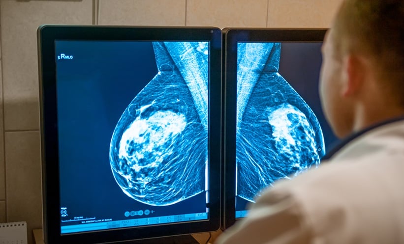
Future research is set to focus on incorporating more detailed patient information and electronic medical record data to further improve diagnostic accuracy. There is hope that similar AI models could be adapted for imaging types other than MRI, including mammograms, chest X-rays and ultrasounds.
Reference
Lyu Y et al. Learning neuroimaging models from health system-scale data. Nat Biomed Eng. 2026;DOI: doi.org/10.1038/s41551-025-01608-0.