PET and MRI scans may together distinguish a new type of dementia from Alzheimer’s disease (AD), a 2026 retrospective, observational study has found.
LATE
Limbic-predominant age-related transactive response DNA-binding protein 43 kDa encephalopathy (LATE), caused by abnormal TDP-43 protein deposits, is emerging as a prevalent neurodegenerative disorder in ageing populations.
LATE mimics the clinical presentation of AD, typically characterised by problems with memory. However, there are different underlying causes, and therefore treatments, for LATE and AD.
Currently, there is no way to diagnose LATE in living people.
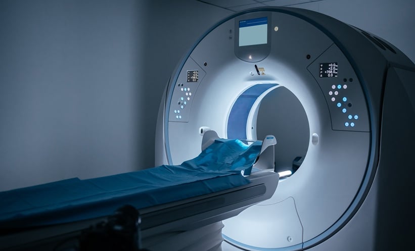
Combining PET and MRI
Researchers carried out a retrospective analysis of nearly 1,000 PET scans from cognitive disorder clinics.
They developed 3-dimensional stereotactic surface projection PET templates for both LATE and AD neuropathologic change, using autopsy-confirmed Alzheimer’s Disease Neuroimaging Initiative and University of Utah datasets.
Researchers then used autopsy-derived z score maps to stratify participants into probable LATE, probable LATE and AD, and probable AD. MRI data was compared across the groups.
Of the PET scans, 13% were characterised as probable LATE (10.6% of which were LATE with AD) and 23.7% were characterised as probable AD without LATE.
MRI Volumetry Findings
MRI volumetry revealed that in pure LATE cases, the medial temporal lobe was most affected.
In mixed LATE and AD cases, the orbitofrontal gyrus and lateral temporal lobe were most vulnerable.
The entorhinal cortex and amygdala proved key regions for distinguishing mixed LATE and AD cases from pure LATE and pure AD cases, respectively.
The same brain hemisphere was normally involved in both LATE and AD cases.
Future Implications
A recognised obstacle to clinical progress in this field is a lack of diagnostic tools, which could supplement studies seeking to define the risk factors and clinical features of LATE.
Clinicians may be able to use MRI and PET scans to detect LATE pathology in patients with cognitive impairment, marking a promising step towards a method for LATE diagnosis.
References
Ngam PI et al. Copathologies of limbic-predominant age-related TDP-43 encephalopathy and Alzheimer disease: [18F]FDG PET statistical mapping and quantitative MRI volumetry. Nucl Med. 2026;DOI:10.2967/jnumed.125.270614.
National Institute of Aging. What is limbic-predominant age-related TDP-43 encephalopathy (LATE)?. 2023. Available at: https://www.nia.nih.gov/health/alzheimers-and-dementia/what-limbic-predominant-age-related-tdp-43-encephalopathy-late#:~:text=On%20this%20page,the%20brain%20and%20other%20tissues. Last accessed: 25 February 2026.
Nelson PT et al. Limbic-predominant age-related TDP-43 encephalopathy (LATE): consensus working group report. Brain. 2019;142(6):1503-1527.
- Author: