Fiona Gilbert | Professor of Radiology, University of Cambridge; AI Lead Advisor, Royal College of Radiologists, London, UK
Citation: EMJ Radiol. 2026;7[1]:57-62. https://doi.org/10.33590/emjradiol/024YY69L
![]()
After graduating from medical school in 1978, what initially motivated you to specialise in radiology, then specifically in breast cancer imaging?
After my house jobs, I did a year in oncology with the late Sir Kenneth Calman, who trained initially as a surgeon and then moved into oncology. He was one of the youngest professors and set up the oncology unit in Glasgow, UK. That year the first CT scanner was installed in Glasgow Royal Infirmary. We sent patients there to decide whether or not they were responding to treatment, and CT scans had only just begun to be used for that purpose.
I remember saying, “We need to see what’s happening here,” and he asked me why, to which I replied, “Because you need the evidence.” At that point, he told me I should be a radiologist. He knew my dad was a radiologist in Glasgow. Initially, I decided against it, because I didn’t want to follow in my father’s footsteps, but I was a visual thinker. I found it much easier to understand something if I could visualise it. When I was learning in medical school, I’d be able to see where something was written on a page. It was not photographic memory, but it was a very visual memory, and I would learn by writing things down, drawing maps, and things like that. I suppose that aptitude for looking at visual things might have been the start.
When I moved to Aberdeen, I completed my Membership of the Royal College of Physicians. I remember saying I would go into public health, or something like that, and the postgraduate Dean said to me, “You’ve got your Membership of the Royal College of Physicians, you passed all the exams, you should be doing something in the hospital.” Therefore, I looked around, and there was radiology training in Aberdeen, so I decided to do that.
You have worked extensively across academic, clinical, and advisory roles. How has maintaining an active clinical practice influenced your research priorities, particularly when assessing which imaging innovations are most meaningful for improving patient outcomes?
It was a piece of advice that my father gave me. Because he knew I wanted to do research, he said, “Don’t give up your clinical work. Make sure that you don’t stop being aware of clinical developments, so you’ve got a really good handle on what’s most important in radiology, and how the field has changed. It means you keep much more abreast of what’s going on and, therefore, it means your research is more likely to be clinically impactful, because you’re working in it on a day-to-day basis.” I still do breast work in the clinic and stay on top of new developments. My main academic theme has been new technology assessment, and what the clinical impact on the patient outcome is. By working in the clinical arena, I can see what’s important, what’s less important, and what the next direction of the field might be.
I’ve been really lucky in my career that there were so many developments going on. CT had just been introduced when I started in radiology training, and that was why we were so excited about it. At the same time, MRI was being developed, and we had a fantastic innovative team of scientists in Aberdeen, where I was working, at the forefront, and that gave me a wonderful opportunity to be really involved at the shop floor level with patients being scanned, and working with the teams there. Fundamentally, they wanted patients to try out their new sequences, and we were able to provide that for them. People talk about being in the right place at the right time. I think, from my career point of view, there was a lot of that. I just happened to recognise the opportunity or be hungry for opportunities at the right times.
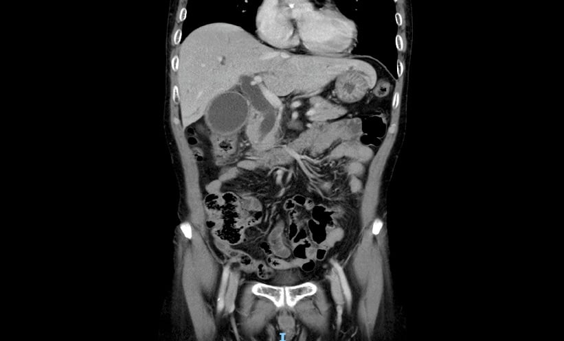
Your research has explored multiple imaging modalities, including tomosynthesis, MRI, contrast-enhanced mammography, and AI-assisted interpretation. Which of these technologies currently offers the greatest potential to improve early cancer detection and diagnostic confidence, and why?
I think all of these technologies have a role to play in terms of early diagnosis. The USA has largely moved to tomosynthesis, which is particularly helpful in women who have got mixed density breast tissue. We’ve completed the PROSPECTS trial in the UK with encouraging results, and I think we will be adopting that technology, at least for some women, and that will increase our cancer detection and reduce our recall rates a little bit, which will be great. However, I think for people who have very dense breast tissue, where we think the mammogram is normal, we should be using AI prediction tools to decide whether or not to offer them additional imaging or bring them back for more imaging a year later. We should be offering them contrast mammography or abbreviated MRI, a fast MRI examination to make sure that we’re not missing a small cancer, to provide earlier diagnosis for those women with dense breast tissue.
There are a lot of different things we should be looking at in the breast screening programme. That is, of course, what makes it difficult for the policymakers, because they don’t know which one to implement. In reality, we need to be pushing all of them. We need to be moving ahead and adopting these different technologies now. I can understand the reluctance just to say, ‘okay, we’re going to move with this’ because, at a population level, you don’t want to be making mistakes. But, in reality, we just need to start doing some feasibility testing in the field at several sites and assessing how it pans out. By putting things in the field, in practice, you learn a huge amount. One of the things we’re doing at the moment is the big EDITH trial, where we’re replacing one of the two human readers with AI for the detection of breast cancer. The good thing about this trial is that we will end up rolling out the AI across about 30 sites in the UK, so that is fantastic. Almost one-third of the UK sites will have exposure to AI in breast imaging.
Overall, I think we need to persuade the people, depending on which healthcare system you’re in, to move to new technology earlier. In some ways, it’s not good enough just to sit on the fence for a long time and wait for yet another trial or health economic analysis.
The BRAID trial, the interim results of which were recently published, aims to demonstrate the value of supplemental imaging in women with dense breasts. Could you describe the rationale behind this trial, and the unmet needs it aims to meet?
In the UK, we do 3-yearly screening, and we’re the only country to have such a long interval between the screening examinations. So, our problem is that we have a lot of interval cancers. A lot of cancers occur in women presenting symptomatically with a lump in their breast or other symptom(s), and women who have interval cancers have a worse outlook compared to smaller screen detected cancers. So, what we’re trying to do is reduce the number of interval cancers and find the cancers a couple of years earlier at the previous screening examination.
The motivation for the BRAID trial was that we knew people who had very dense breast tissue had more interval cancers and had larger cancers, so we thought, let’s target these women with an additional imaging technique. The question we are trying to answer is which of the imaging techniques was better? Is a fast MRI scan better than a contrast mammogram or whole breast ultrasound? Contrast mammogram is very effective for showing these small cancers. Cancers have lots of blood vessels that take up the iodine and show up brightly on the mammogram, which is fantastic. An MRI, we’ve known for a long time, is much more sensitive than any of the other techniques, but it’s quite costly. A fast MRI scan reduces the cost by about 50%, so we wanted to compare contrast mammography with fast MRI against the standard supplemental imaging, which is breast ultrasound. That was what the trial was about.
We recruited over 9,000 people across 10 sites in the UK, and randomised them to one arm each. We did the additional imaging and then did a second round of imaging, and we found that the two contrast techniques, abbreviated MRI and contrast mammography, picked up between 16–18 additional cancers per 1,000 women that we screened, compared to ultrasound, which only picked up four additional cancers. We’re seeing as many as four-times as many cancers in the contrast arms compared to the ultrasound arm. This was a very exciting study, and we felt that this was definitive evidence that you should be doing contrast techniques, not ultrasound, which is offered in other countries, such as the USA, Italy, and Germany, to women with dense breasts, but, actually, it’s not good enough. We should be doing these other tests instead.
The interim results from BRAID suggest significant benefits of supplemental imaging for women with dense breasts. What are your expectations for the full results when they are released in a few years’ time? And how are these set to impact patient outcomes?
What we’re seeing with the second-round imaging is another eight cancers per 1,000, which is amazing, because we’re only imaging 18 months later. So, if you think about it, in the Breast Imaging-Reporting and Data System (BI-RADS) D category, we’ve already found eight cancers per 1,000 from the mammogram. So, if you take women with a negative mammogram and offer them contrast examination, you pick up another 18 cancers per 1,000. Then, 18 months later, you do another contrast examination, and you pick up another eight cancers per 1,000. That’s 34 cancers per 1,000 in that 18-month period, which is amazing, but you think, where are all these cancers coming from? And that’s why we have to wait for the results of the main study, because we need to look at the number of interval cancers to see if there is a reduction in the interval cancers too. Also, we need to look and see what the next round of mammography cancers are. We’d expect to see a dip in the cancer detection rate of the next round, and certainly a smaller tumour size at the 3-year mark as well, because we’ve brought the diagnosis forward for so many of these women.
You have been closely involved in the development and evaluation of AI tools for breast imaging, and you are now the Lead AI Advisor at the Royal College of Radiologists. As AI becomes increasingly integrated into clinical practice, where do you see its greatest potential in breast imaging? Are there particular areas where you believe it will have the most meaningful clinical impact?
AI is moving at an extraordinary pace. In the UK, we’re still in the relatively early stages of its widespread adoption, but imaging is leading the way. One of the reasons for that is that radiology has long worked with digital images, providing exactly the kind of structured data that AI systems can be developed and trained on. I think the key is that we’re going to get improved standardisation of reporting quality. What previous work has shown is that people who are less confident and less experienced often show an improvement in their performance when they’re using AI tools, with prompts, etc., whereas, for the more experienced readers, we often don’t see much of a change. While not everybody can be seen by the best musculoskeletal radiologist in the country, often the AI can help lift many people up towards that level, which I think is one of the important things.
Secondly, I think it’s going to make us monitor the performance of the AI and look at human interaction with the AI. In breast work, we’ve been very used to regular quality assurance and performance monitoring (how many cancers do I detect a year? How many cases do I recall from screening?). There are a number of metrics on which I’m measured. I think that kind of audit will become more mainstream across radiology.
I also think, quite shortly, we’re going to see a shift to autonomous reporting of high-confidence normal examinations. About 30% of all our examinations are normal. They are the people who are referred because we need to exclude ‘X, Y, and Z’, and we end up with a normal chest X-ray or normal CT. We already know that when you look at the chest X-ray algorithms, it’s trying to pick up around 20 different conditions. If it’s a high-confidence normal examination, then it is genuinely normal. When you compare that with a whole group of different reporters, then you actually get more accurate results with the AI than you do with this variable group of people that are reporting the chest X-ray. So, I think we will see a shift towards AI being used for this purpose in very controlled circumstances, because we need to get the public to understand what we’re trying to do. We’re not trying to save money. We’re trying to free up capacity so that we can deliver a better radiology service in a more timely way and do better for patients. We know that radiologists are going to be doing more complex imaging and reporting, but I think we’re going to be able to use the AI tools in some areas for cases that are genuinely normal. It’ll be able to cut down maybe 30% of the normal reporting because the AI tools will be sufficiently confident to say this is definitely normal, and the miss rate would be much less than a human reporter. It is very important that we improve the delivery of radiology services by having more radiologists to match the 8% year on year rise in imaging tests, and AI will help us deliver radiology services more efficiently. Clinicians are increasingly dependent on imaging: like me, they want to “see” what is going on. I think that’s a better service for the public, which is why I think that is one of the shifts we are going to see.
A very important aspect of introducing a new system such as AI is patient safety. It is essential to audit the performance of AI and the human interaction. However, with uncommon errors, a centralised reporting system could be helpful, so that if there is an adverse event in one hospital, it can be reported, and patterns of adverse events can be recognised more quickly around the country. In summary, if a trust has one adverse event in a year, then they might not think anything of it, but if we see the same adverse events from 20 different hospitals, that needs to be collated and something needs to be acted on quickly. That’s what the Medicines and Healthcare Products Regulatory Agency (MHRA) are looking at; I think they are looking at strengthening their yellow card system, which is used mainly for drug and medical device reporting currently.
Despite strong evidence for emerging imaging technologies, adoption into routine clinical practice can be slow. From your experience, what are the most significant barriers, whether technical, organisational, or educational, to implementing innovations, such as AI or supplemental screening?
There are a number of things that need to be put in place. While several trusts have implemented AI for chest X-rays, we still need to encourage trusts to embrace AI technology. Our IT departments and governance are still quite apprehensive about it, so we need to do things in terms of reassuring different areas of the workforce that these tools are safe. I think there needs to be some kind of centralised system whereby we can have shared documents and risk assessments for AI tools, which can help protect identifiable patient information in a trust, as each trust is legally responsible for patient data. That shouldn’t change; that is sacrosanct, and trusts get very nervous when an AI tool is introduced and is scurrying around all the identifiable data. So, we need to harmonise these Data Protection Impact Assessment (DPIA) documents, which provide a reassurance to the trust, and make sure that, however it’s being used, their data is going to be safe.
There needs to be some kind of national accreditation or national approval body, which says, ‘yes, this is good to go’. Then, the trust might not have to spend so much time scrutinising whatever the particular tool is and confirming whether it works in a UK population. There are no ethnic biases or other biases that we should be worried about. The information governance is being properly considered, and that means it can be rolled out more quickly across trusts to streamline that deployment.
Then, we need to be very clear about how we’re going to monitor these tools. We need to make sure that trusts have generic tools in place so that they can just press a button on one of their dashboards and confirm a tool is performing correctly, basically auditing it. This does not need to be a continuous monitoring system, but there should definitely be a monitoring system in place, running in the background, so that a trust can easily pull out data to say the performance is not drifting, the radiologists are still reporting the correct number of abnormals, and so on.
There is quite a lot of work to do, but I think people are aware of that, and of course, it’s upskilling the workforce too, so that they feel comfortable in using AI and understand the limitations of a particular tool. For example, so they don’t start using a tool that’s designed for adults in a paediatric population.
Equity is an important consideration in imaging across geographical areas, with variation in access to advanced imaging remaining a challenge. What practical steps can/should healthcare organisations take to ensure more equitable access to improved screening and diagnostic tools?
I think policymakers are really focussed on this, and they want to do whatever they can to ensure that everybody’s invited, that we look and see why people don’t come for imaging early, why they don’t see the general practitioner (GP), and why they don’t access their screening. There is a lot of work going on to understand what happens in different demographic patterns, different geographic areas, different ethnicities. There are all kinds of things that influence behaviour, so it’s very much an important topic to always consider, particularly when we’re looking at changing access to a system. Although many people have mobile phones and can receive text messages, it’s still not universal, and emails and online activity can be quite a barrier for some people, so we have to always ensure that people can be supported, whether it’s through their GP, which is often a good mechanism to do so, or otherwise. People say, ‘they could go to a library and access online material there’, but I think people are reluctant to go and consider their private healthcare matters unless it is with someone appropriate. I think GPs have a big role to play in ensuring equity of access to all kinds of imaging and screening services. We also need to do what we can to support the GPs. It’s not fair just to shift the weight onto them. We need to put tools in their hands to allow for improved access. We need to make it as easy for the GPs as possible.
For healthcare professionals working in breast cancer today (and those of the future), what practical steps can they take to ensure they are delivering the highest standard of care to their patients?
The amazing thing that’s happened with breast cancer, apart from all the imaging and the incredible technological developments, is that the drugs are so much better. Now that the treatments are so much better, we can minimise treatments and ensure that the right person receives the right treatment for them; advances in personalised medicine are amazing.
I think for people coming into breast imaging, it’s really important to be hopeful about the specialty. I think our role is going to change slightly over time, because we’re going to see more and more people who are living with breast cancer. We’re going to be seeing more people with metastatic disease living longer, so I think we need to make sure that we are very familiar with the different techniques and also interventional techniques, such as vacuum excisions using image guidance and cryotherapy, which are fantastic. There are all kinds of developments that are coming our way, which are presenting amazing opportunities for radiology. It’s fantastic. The future of the specialty of radiology is very bright.