Abstract
Cardiac imaging is crucial for diagnosing and managing coronary artery disease. While most imaging modalities focus on either anatomical or physiological assessment of coronary artery disease, novel hybrid techniques capable of providing both in a single imaging session represent an important advancement in the field. This review summarises the technique, diagnostic performance, clinical utility, limitations, and cost-effectiveness of the principal hybrid modalities: coronary CT angiography (CCTA) c, PET with CCTA (PET-CCTA), and cardiovascular magnetic resonance (CMR) imaging. CCTA with fractional flow reserve applies computational fluid dynamics modelling to standard CCTA data to derive non-invasive fractional flow reserve estimates without additional radiation or contrast exposure. Diagnostic accuracy, at appropriate values, can be high enough to reduce rates of invasive coronary angiography. The application of AI to CCTA for automated plaque quantification and stenosis grading is promising but currently limited by its discordance with invasive angiography. PET-CCTA combines myocardial perfusion imaging with coronary anatomy in a single session with higher specificity than CCTA alone, but at the cost of greater radiation exposure. Finally, CMR provides radiation-free tissue characterisation across multiple techniques such as stress perfusion, late gadolinium enhancement, and strain imaging, each with their unique features. Taken together, PET-CCTA currently offers the most robust integration of anatomy and physiology, while CMR continues to advance rapidly as a compelling alternative, particularly where radiation-free evaluation is preferred. The authors have summarised key evidence on how these hybrid approaches enhance detection of obstructive coronary stenosis, guide therapeutic decisions, and how these technologies are implemented today.
Key Points
1. Non-invasive cardiac imaging modalities typically address either anatomical or physiological assessment of coronary artery disease. Emerging hybrid techniques address both in a single imaging session, potentially reducing unnecessary invasive procedures.
2. This narrative review evaluates the diagnostic performance, clinical utility, limitations, and cost-effectiveness of coronary CT angiography (CCTA) with fractional flow reserve, PET-CCTA, and cardiovascular magnetic resonance imaging as hybrid approaches integrating coronary anatomy with functional assessment.
3. PET-CCTA currently offers the most robust combined anatomical–physiological assessment, while cardiovascular magnetic resonance imaging provides compelling radiation-free alternatives. Clinicians should tailor modality selection to patient-specific factors and clinical context.
INTRODUCTION
Non-invasive cardiac diagnostic imaging is an integral component of coronary artery disease (CAD) identification, risk stratification, and disease management. The anatomic and physiologic information derived from the different imaging modalities shapes the disease and treatment course.1 Most imaging techniques excel at either anatomic or physiologic information about CAD. CT is excellent for assessing anatomic changes of the structures of the heart, and coronary CT angiography (CCTA) has become one of the most frequently used non-invasive imaging modalities to diagnose CAD, specifically in patients presenting with angina with no history of CAD.1 In contrast, PET is limited in its ability to provide anatomic data but excels at assessing physiologic information.2 Cardiovascular magnetic resonance (CMR) imaging and its various techniques can provide both structural and functional information.3
Both anatomic and physiologic variables are valuable in making clinical decisions, and novel hybrid techniques have been developed to provide both in a single non-invasive imaging test. Examples include CCTA with fractional flow reserve (CCTA-FFR) and cardiac PET with CCTA (PET-CCTA). Advances in CMR imaging techniques have also allowed for improvements in coronary magnetic resonance angiography (CMRA), CMR with strain imaging, CMR–PET, and more to offer similar anatomic and physiologic data. Blending anatomic and physiologic imaging techniques can potentially expedite risk stratification, enhance clinical understanding, and reduce unnecessary invasive procedures.4 Recent trials have demonstrated that many patients with stable coronary disease can be managed conservatively, helping clinicians identify patients who can be safely treated without invasive procedures.5 This review provides an overview of the current hybrid cardiac imaging modalities and discusses their techniques, advantages and disadvantages, outcomes, utility, and cost in the diagnosis and management of CAD.
CARDIAC CT
CCTA involves intravenous administration of contrast to enhance visualisation of the coronary arteries during a high-resolution, ECG-gated CT scan. According to the 2021 guidelines from the American Heart Association (AHA) and the American College of Cardiology (ACC), CCTA has Class I-A evidence in intermediate-risk patients with acute chest pain and no known CAD, and Class IIa-B non-randomised evidence in patients with inconclusive or abnormal stress testing.6 Because of its high negative predictive value for ruling out significant disease, defined as >70% luminal stenosis in at least one major coronary artery or >50% stenosis in the left main coronary artery, the use of CCTA in the diagnostic and management pathways of CAD is increasing.1,6 Two pivotal randomised trials have since provided long-term outcome data reinforcing CCTA’s value beyond its established rule-out capability. In the PROMISE trial (N=10,003; median follow-up 10.6 years), initial test selection did not affect mortality, but any CCTA abnormality, including non-obstructive disease, conferred elevated long-term risk (adjusted hazard ratio: 1.99–3.44), whereas only severely abnormal stress results were prognostically significant.7 The SCOT-HEART trial (N=4,146; 10-year follow-up) demonstrated that adding CCTA to standard care reduced coronary heart disease death or non-fatal myocardial infarction (hazard ratio: 0.79; 95% CI: 0.63–0.99; p=0.044), driven by increased preventive therapy use without excess revascularisation.8 These findings underscore that CCTA’s detection of atherosclerosis provides a management-relevant anatomic foundation upon which hybrid physiologic assessments can build.
CCTA-FFR
CCTA-FFR is an adjunct to CCTA that provides FFR measurements calculated through computational fluid dynamics modelling algorithms, with the goal of non-invasively determining the severity of coronary stenosis.9-11 CCTA-FFR is an add-on test that does not require additional exposure to radiation or administration of contrast agents beyond those used for acquisition of the original CCTA.11 It uses advanced computational models to build a 3D, patient-specific model of coronary anatomy and provide FFR estimates for each lesion. These models are calibrated against invasive coronary angiography (ICA) FFR, in which values ≥0.80 can rule out ischaemia.12
In a systematic review published in 2017, the authors concluded that the diagnostic accuracy of CCTA-FFR varies markedly across the spectrum of CAD. Accuracy was highest when lesions were identified with CCTA-FFR values lower than 0.63 or above 0.83, but lower with values between 0.63–0.83, where FFR is most needed to help rule in or out ischaemic lesions.13 Ultimately, the authors suggested that by using CCTA-FFR in combination with patient-specific factors, clinicians may be able to identify cases in which cost and risk of an ICA may safely be avoided. Other trials and systematic reviews have offered similar findings and conclusions for CCTA-FFR to be used as an adjunct to clinical acumen.14-16 Limitations of CCTA-FFR are similar to standard CCTA: motion artefact, obstruction to the lumen by coronary artery calcification, use of iodinated contrast, and ionising radiation.17 Coronary calcification is especially prevalent in patients with longstanding chronic kidney disease or diabetes, in whom heavy vascular calcification frequently produces blooming artefacts that obscure luminal assessment and reduce CCTA diagnostic accuracy.17 CCTA-FFR-specific limitations include reduced validity in patients with coronary stents, bypass grafts, coronary anomalies, transcatheter aortic valve replacement, and more.4,17
CCTA-FFR Discussion
A brief note regarding limitations in CCTA-FFR: emerging photon-counting detector CT offers improved spatial and contrast resolution and reduced artefact, with particular advantages for evaluating heavily calcified vessels and small-calibre stents. In a comparative study, photon-counting detector CT demonstrated significantly higher diagnostic accuracy than conventional CT for detection of >50% and >70% stenosis at the patient, vessel, and segment level, reducing use of ICA.18
When it comes to financial implications, the PLATFORM trial found that when CCTA-FFR was used prior to percutaneous coronary intervention in patients with stable, new onset chest pain, total healthcare costs were 33% lower at 1 year.19 The UK-based FORECAST trial and its USA cost analysis found that CCTA-FFR did not significantly reduce overall cardiovascular care costs at 9 months compared to CCTA or stress imaging alone, though it did reduce the rate of ICA.20,21 Another UK-based study showed that CCTA-FFR was slightly more expensive than CCTA alone, with a mean per-patient increase of 44.97 GBP, but it accelerated time to definitive diagnosis and reduced unnecessary invasive procedures.22 Although there may be slightly higher upfront costs with CCTA-FFR depending on the healthcare system, reducing the rate of invasive procedures may decrease overall healthcare costs in the long term.
AI in Cardiac CT
Beyond computational modelling, AI techniques have emerging applications in CCTA, particularly in image acquisition, reconstruction, and interpretation.23,24 The REVEALPLAQUE study applied an AI algorithm to compare plaque assessment on CCTA with intravascular ultrasound (IVUS).25 Clinically stable patients with known CAD underwent CCTA-FFR, followed by ICA and IVUS. CCTA images were processed with an AI algorithm, which calculated total plaque volume, lumen volume, vessel volume, calcified plaque volume, and non-calcified plaque volume. Compared with IVUS, the system performed well regarding its primary endpoint of total plaque volume and secondary endpoints, including calcified plaque volume, non-calcified plaque volume, lumen volume, and vessel volume (r=0.91, 0.91, 0.87, 0.93, and 0.94, respectively; p<0.001 for all). However, performance was weaker for low-attenuation plaques (r=0.28). The spatial resolution of CCTA (approximately 0.5 mm) is lower than that of IVUS (approximately 0.04–0.20 mm), which may reduce sensitivity for detecting small or thin low attenuation plaques, particularly in smaller vessels. A CREDENCE trial substudy compared AI-enhanced CCTA to ICA.26 While the authors of this study are confident in their results showing similar accuracy between AI-enhanced CCTA and ICA, there were significant inconsistencies identified. False positives occurred when AI-enhanced CCTA overestimated stenosis due to high calcification or poor lumen opacification. False negatives occurred when AI-enhanced CCTA detected significant stenosis that ICA did not confirm. Among 157 vessels classified as having ≥70% stenosis by AI-enhanced CCTA, 62 cases (39.4%) were discordant with ICA findings. Notably, 41 of these discordant cases demonstrated FFR <0.8, suggesting that AI-enhanced CCTA may capture physiologic significance that traditional ICA underestimates, due to its 2D assessment compared with 3D AI-based analysis. However, the authors did not address diagnostic and management challenges in the remaining 21 cases where AI detected significant stenosis without true functional lesions (patients who may be exposed to unnecessary invasive testing).
AI in Cardiac CT Discussion
Given the current limitations of AI-enhanced CCTA, it remains unclear whether this technology should be routinely incorporated into the diagnosis and management of CAD.
Present models lack precision in evaluating lesions, particularly low-attenuation plaque. The degree of discordance between AI and physician interpretation remains substantial and may result in ambiguous findings that lead to unnecessary invasive testing on an individual basis. Nonetheless, existing studies underscore that, while AI-enabled CCTA is not yet perfected, it is an active area of research and a promising avenue for future growth.
With continued advancements, AI models are expected to support physicians more effectively, narrowing the gap between computer- and physician-read CCTA. Its potential cost-effectiveness will depend largely on the ability to reliably rule out ischaemia-provoking CAD and thereby reduce the need for downstream invasive testing, such as ICA.27
PET with Cardiac CT
PET myocardial perfusion imaging uses radiotracers to provide detailed information on myocardial perfusion and metabolic activity, which is crucial for evaluating the functional significance of CAD.2 It is particularly useful for detecting ischaemia and assessing myocardial viability. Diagnosing CAD via PET has sensitivities of 90–94% and specificities of 79–90%.28 PET can be performed as a myocardial perfusion study alone or in conjunction with coronary flow reserve (CFR). PET-derived CFR is a quantitative, non-invasive measure of the ratio between maximal (hyperaemic or stress) and resting myocardial blood flow, obtained using PET tracers administered during the scan.29,30 CFR is measured by acquiring dynamic PET images at rest and during pharmacologic vasodilation, followed by kinetic modelling to quantify the coronary circulation’s ability to augment blood flow in response to increased myocardial demand. When applied to obstructive CAD, this technique has demonstrated a sensitivity of 90% and a specificity of 88%.31
PET-CCTA Hybrid Imaging
The novel PET-CCTA hybrid imaging modality combines PET myocardial perfusion imaging and CCTA in a single session, providing simultaneous anatomical and physiological assessment of CAD.32 This integration theoretically enhances the diagnostic accuracy for non-invasively detecting CAD, evaluating tissue ischaemia in the same imaging session, and maintaining the ‘rule out’ feature of CCTA alone.33-35 The process of capturing PET-CCTA images starts with CCTA performed first, followed by PET, often using radiotracers (most commonly rubidium-82 [82Rb] or 13N-ammonia [13NH3]) and pharmacologic stressors, such as regadenoson.36 Fused comprehensive images show both anatomical and physiological information, allowing direct correlation of perfusion defects with specific coronary lesions. A 2018 meta-analysis compared hybrid imaging to CCTA for the diagnosis of obstructive CAD. It showed that PET-CCTA had a sensitivity and specificity of 87% and 96%, respectively, compared to 90% and 66% for CCTA alone.37 While the change in sensitivity was not statistically significant, the improvement in specificity was p<0.05. The authors discuss that the increased specificity makes PET-CCTA and other hybrid imaging modalities efficient, non-invasive diagnostic tools to help rule in lesions that may be intervenable and avoid targeting non-functionally limiting lesions. Similar findings were reported in a prospective study of 208 patients that compared multiple non-invasive imaging modalities against invasive FFR as the reference standard.38 PET-CCTA improved specificity to 92% compared with 84% for PET alone, but decreased sensitivity to 74% from 87%. The authors attributed the reduction in sensitivity largely to the small sample size. Nonetheless, the higher specificity supports PET-CCTA’s ability to identify lesions appropriate for invasive targeting based on functional as well as anatomical data.
PET-CCTA Discussion
Given the mixed evidence regarding sensitivity, CCTA alone remains a reasonable option for low-risk patients without prior CAD when the clinical aim is to rule out disease. However, PET-CCTA may offer particular value in patients with known non-obstructive CAD who present with new or progressive symptoms, or in cases where CCTA is limited, such as heavily calcified lesions that are frequently overestimated. Limitations of PET-CCTA include higher radiation exposure compared with CCTA alone, owing to the combined CT and PET components.39 The use of iodinated contrast agents also poses risks in patients with severe renal dysfunction or contrast allergy.6 Accessibility remains a challenge, as PET-CCTA requires specialised hybrid scanners and radiotracer supply (e.g., on-site cyclotron for 13NH3 or generator for 82Rb), which are not universally available. Cost is another barrier: patients utilising in-network radiology providers pay on average 319 USD for PET myocardial perfusion studies, with some estimates exceeding 1,000 USD.40 Cost analyses directly comparing PET-CCTA with other non-invasive modalities are limited, though it is anticipated to be more expensive than PET alone.
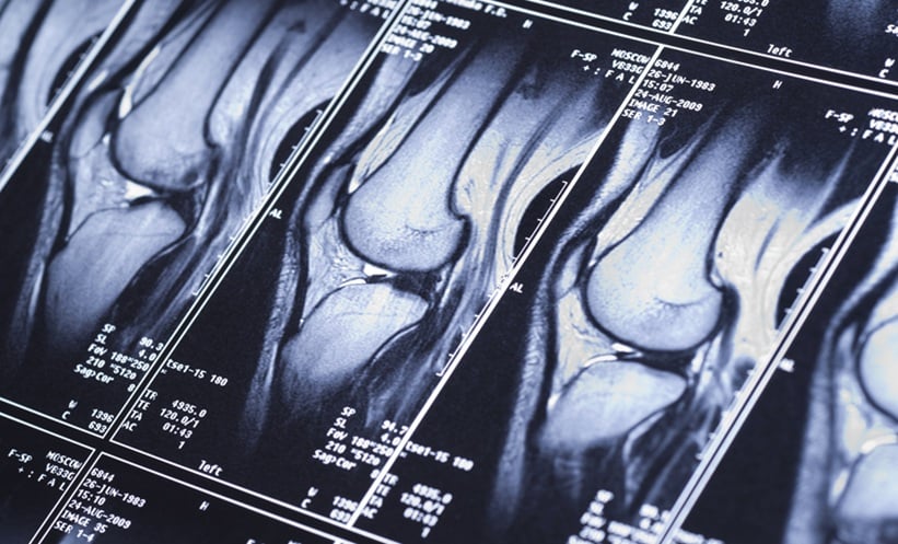
CMR IMAGING
CMR imaging allows assessment of cardiac function, ischaemia, viability, and tissue characterisation within a single scan.41 Over the past decade, numerous studies have established its role in stable CAD, contributing important technical advances, large-scale clinical validation, and prognostic data. As a result, CMR has emerged as a valuable tool for diagnosis and risk stratification in CAD.6,31 CMR offers several advantages over other imaging modalities. It is particularly useful in patients where minimising radiation exposure is important, such as younger individuals or pregnant women. Compared with nuclear imaging, CMR provides superior spatial resolution, and, unlike echocardiography, it is not limited by acoustic window constraints.42 It also provides better temporal resolution than cardiac CT.43 These characteristics make it a strong modality for evaluating ventricular anatomy and function, including wall motion abnormalities.
Stress Perfusion CMR
Stress perfusion CMR is unique in its ability to directly visualise the myocardium during stress. Its high spatial resolution allows comparative assessment both between myocardial segments and between the epicardium and endocardium. Tissue characterisation with T1, T2, late gadolinium enhancement (LGE), and blood oxygen level-dependent imaging enables identification of scar, haemorrhage, oedema, and regional hypoxaemia.
Multiple meta-analyses and prospective multicentre studies report a sensitivity of 78.9–91.0%, a specificity of 81.0–86.8%, and an area under the curve of 0.84–0.87 for detecting significant CAD, defined as ≥70% stenosis by ICA or FFR. Negative predictive value is high, with 1-year event rates for cardiovascular death <1% following a negative test.31,44-48 Another technique, CMRA, visualises the coronary arteries directly using MRI.49,50 CMRA can also provide information on coronary artery distensibility, plaque characteristics, and plaque inflammation.50 Depending on the protocol, vasodilators, inotropes, or exercise are used to evaluate myocardial blood flow under stress, followed by administration of a gadolinium-based contrast agent to assess myocardial scar and viability.51 According to the American College of Radiology (ACR), non-contract CMRA is most useful for visualising the coronary tree in symptomatic patients with intermediate or high pretest probability, particularly for ruling out significant CAD. Its diagnostic yield is further enhanced when combined with stress perfusion and/or LGE CMR protocols, which add functional information.31 CMRA alone can identify significant coronary artery stenoses, with recent multicentre studies demonstrating a sensitivity of 88–96% and a negative predictive value of up to 93% for detecting ≥50% stenosis. However, specificity and positive predictive value remain more modest (typically 68–79%).31,52,53 Despite these advances, CMRA remains technically challenging to perform.
Late Gadolinium Enhancement CMR
LGE CMR is highly accurate for detecting significant CAD, demonstrating a sensitivity of 89%, a specificity of 94% in patients with new left ventricle dysfunction, and diagnostic accuracy of 92%.54 LGE is also a strong independent predictor of adverse cardiac events.55 When combined with stress perfusion imaging, LGE provides complementary diagnostic and prognostic information, with the absence of both findings conferring a very high negative predictive value for major adverse cardiac events.56 CMR strain imaging is another promising non-invasive modality. Strain CMR refers to the quantitative assessment of myocardial deformation using computational models, such as feature tracking, strain-encoded imaging, myocardial tagging, or displacement encoding with stimulated echoes.57,58 These methods quantify myocardial fibre shortening or lengthening in longitudinal, circumferential, and radial directions, providing a sensitive marker of subclinical myocardial dysfunction that often precedes reductions in ejection fraction or echocardiographic evidence of wall motion abnormalities.58,59 CMR strain imaging is particularly useful in patients requiring detailed quantification of ischaemic, non-ischaemic, or valvular heart disease. Both left ventricular long-axis measurements and circumferential or longitudinal strain values are expressed as negatives, with thresholds greater than –17% and –20% considered pathologic, respectively.58
CMR Strain Imaging
In the context of CAD, CMR strain imaging is valuable for both diagnosis and risk stratification. Strain analysis can detect regional and global myocardial dysfunction due to ischaemia or infarction, even when conventional markers, such as ECG, troponin, and ejection fraction, remain preserved. In a study of 108 patients, fast strain encoded CMR and feature tracking CMR demonstrated a sensitivity of 82%, a specificity of 87%, and a negative predictive value of 96% for flow-limiting CAD confirmed by ICA in patients presenting with acute chest pain.60 The authors concluded that CMR strain outperformed traditional markers, such as ECG and troponin dynamics, providing a high negative predictive value for ruling out flow-limiting CAD.
For prognostication, reduced global or regional strain measured by CMR is independently associated with major adverse cardiac events in patients with known or suspected CAD.61 Recent studies show that stress CMR with strain analysis, particularly global circumferential strain, adds incremental prognostic value to conventional CMR parameters and can predict cardiac death, nonfatal myocardial infarction, and heart failure hospitalisation.61,62 Notably, CMR strain assessment has been reported to provide prognostic utility comparable to CMR stress perfusion imaging, which is especially relevant for patients with contraindications to gadolinium.62 CMR strain analysis has also been effective in measuring myocyte recovery following cardiac injury.58
CMR-PET
CMR-PET is a hybrid imaging modality that combines the high spatial and tissue characterisation capabilities of CMR with the molecular and quantitative perfusion imaging of PET.63-65 This approach enables simultaneous or sequential acquisition of anatomical, functional, and metabolic data, offering comprehensive assessment of coronary artery anatomy, myocardial tissue characteristics, blood flow quantification, and detection of inflammation or metabolic activity. A 2024 review article highlighted recent advances in CMR-PET technology.63 A small portion of their review discussed the utility of this hybrid imaging as it relates to myocardial perfusion. With respect to CAD, only limited data exist: in a small study of 15 patients, Kero et al.66 assessed 15O-water myocardial blood flow using CMR-PET and reported good correlation and moderate agreement with simultaneous dynamic contrast-enhanced MR assessments. To date, no large-scale, prospective studies or meta-analyses have directly compared CMR-PET with other modalities for CAD diagnosis.
CMR Discussion
Despite its strengths, CMR is limited in certain patient populations depending on the protocol. It is contraindicated in patients with non-MRI-conditional implanted devices and requires caution in those with severe renal dysfunction, due to the risk of nephrogenic systemic fibrosis with gadolinium, although many indications do not require contrast. Severe claustrophobia can also pose challenges, though newer open-bore and upright scanners are improving accessibility. Patient participation, such as breath-holding, may also be limiting.6 Compared with CCTA, CMR has lower spatial resolution (2.0 mm versus 0.4–0.6 mm), significantly longer acquisition times (up to 1 hour), and more complex planning requirements.49 These remain barriers to widespread adoption, though advances in acquisition speed are improving tolerability. Careful patient selection and consideration of these limitations remain essential when choosing CMR for CAD evaluation.
Cost-effectiveness has also been studied. The 2021 AHA and ACC guidelines on chest pain cite UK-based studies, showing that CMR strategies are more cost-effective than single-photon emission CT, CCTA, or immediate ICA in patients with low-to-intermediate pretest probability of CAD, with comparable or lower costs.6 The inherent redundancy in CMR data acquisition allows cross-verification of findings, for example, the same myocardial segment can be assessed in short-axis and long-axis cine images, as well as through perfusion and tissue characterisation, helping mitigate incomplete coverage, motion artefacts, or segmentation errors.67 CMR is particularly valuable in evaluating conditions that mimic or coexist with CAD, such as myocarditis, takotsubo cardiomyopathy, or myocardial infarction with non-obstructed coronary arteries.6 In the USA, a cost analysis using data from the Stress CMR Perfusion Imaging in the United States (SPINS) registry found that a CMR-first strategy can serve as a cost-effective gatekeeper prior to invasive angiography in patients with stable chest pain syndromes.68 However, additional USA-based cost-effectiveness data comparing CMR with other non-invasive modalities remain limited.
CONCLUSION
Among the available modalities, PET-CCTA currently provides the most robust integration of anatomy and physiology, offering improved specificity for identifying functionally significant lesions and guiding invasive management when appropriate. CCTA remains the first-line anatomic test, due to accessibility and its ability to exclude CAD, with AI and FFR CT promising to strengthen its physiologic relevance. CMR adds unique value through radiation-free tissue characterisation and emerging strain techniques, making it an ideal complement in select populations. Taken together, while each modality has a role, hybrid PET–CCTA best exemplifies the combined anatomic–physiologic approach today, with CMR advancing quickly as a strong contender.