SUPRACARDIAC atherosclerosis, which affects arteries in the head, neck, and aortic arch, is a major contributor to ischaemic stroke. Current imaging techniques such as CT angiography (CTA) provide efficient coverage but involve radiation exposure and contrast agents. A new vessel wall MRI (vwMRI) protocol promises improved plaque detection, providing clinicians a more detailed, non-invasive assessment in a clinically feasible timeframe.
Understanding Supracardiac Atherosclerosis and Stroke Risk
Many patients with embolic stroke of undetermined source (ESUS) have plaques in supracardiac arteries, sometimes accompanied by complex aortic plaques, pointing to a wider vascular source of the stroke. According to this study, ipsilateral non-stenotic plaques appear in approximately 40% of ESUS cases, and around a third of these patients exhibit complex aortic plaques, highlighting a systemic origin of emboli. Multivascular disease further increases the risk of recurrent stroke by about 1.5 times compared with single-territory atherosclerosis.
Study Design and Imaging Innovations
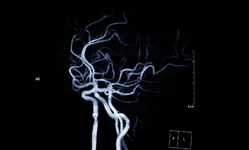
Researchers developed a non-contrast head-neck-aortic vwMRI integrating multichannel coils, grey-blood imaging, and neural network-based acceleration, achieving whole supracardiac coverage in around 15 minutes. Between October 2024 and March 2025, 108 participants with a history of ischaemic stroke or transient ischaemic attack were prospectively enrolled at a tertiary centre in Beijing to undergo both routine CTA and the new vwMRI protocol. Plaque features including calcification, ulceration, and intraplaque haemorrhage were compared, and CTA served as the reference for calcification assessment.
Improved Detection and Reclassification of Stroke Aetiology
The vwMRI protocol identified plaques in 815% of participants versus 69.4% with CTA and achieved 91% accuracy in detecting calcifications. Intraplaque haemorrhage was detected in 27.8% of patients, a marker of plaque vulnerability that CTA often misses. Ulceration rates were comparable between modalities. Notably, among 38 patients initially classified with ESUS via CTA, vwMRI enabled reclassification in 16 cases, reducing undetermined stroke cases from 35.2% to 20.4%.
Limitations and Considerations
This study lacked histopathologic validation of imaging findings and excluded patients who could not undergo MRI, potentially introducing selection bias. The single-centre design and modest sample size, and overwhelmingly male cohort (89 men, 19 women) also limit generalisability, highlighting the need for multicentre studies with more balanced participant demographics to confirm efficacy and workflow integration.
Implications for Clinical Practice
By combining rapid acquisition with detailed vessel wall assessment, this vwMRI protocol offers a radiation-free alternative to CTA, capable of detecting vulnerable plaques and refining stroke aetiology determination. Its 15-minute scan balances speed with comprehensive coverage, presenting a practical tool for clinicians managing patients at risk of recurrent embolic events. This approach may ultimately streamline stroke workflows and support more targeted preventative strategies.
Reference
Jiang Q et al. Evaluation of Supracardiac Atherosclerosis in Stroke with a Noncontrast Head-Neck-Aortic Vessel Wall MRI. Radiology. 2026;DOI:10.1148/radiol.251586.
Featured image: Samunella on Adobe stock
- Author: