LEFT VENTRICULAR myocardial reference values derived from cardiac CT feature tracking show clear sex-based differences in healthy adults, offering clinicians a clearer benchmark for interpreting cardiac imaging.1
Myocardial strain, a measure of how heart muscle fibres deform during contraction and relaxation, is a sensitive indicator of myocardial dysfunction and may detect abnormalities earlier than conventional measures such as ventricular ejection fraction.2 Establishing normal reference ranges is therefore important for clinical assessment.
Why Left Ventricular Myocardial Strain Matters in Practice
Left ventricular myocardial strain reflects the mechanical performance of the heart and is assessed across three dimensions: longitudinal, circumferential and radial strain. These measures provide additional insight into cardiac function, particularly in patients with subtle or early-stage disease where ejection fraction may still appear normal.2
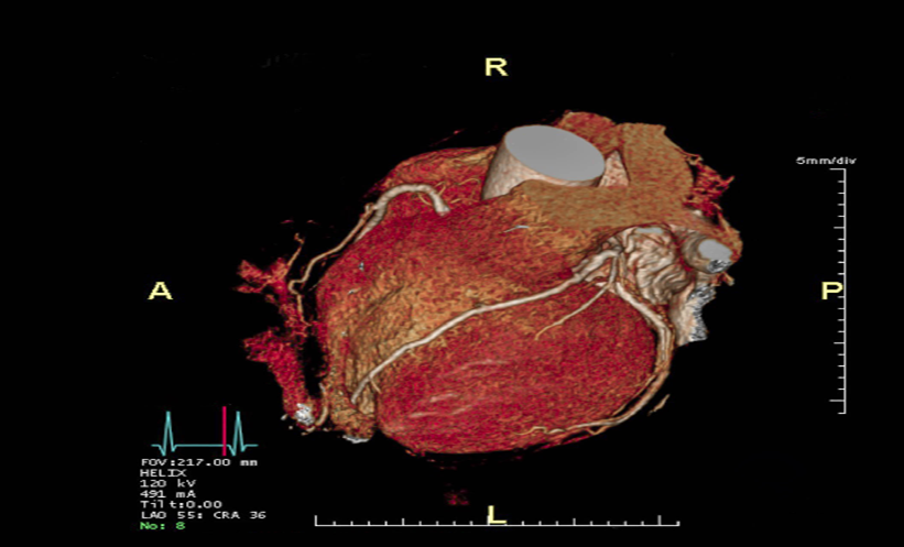
This study determined reference values for left ventricular myocardial strain using CT feature tracking in healthy participants undergoing coronary CT angiography.1
CT-Based Left Ventricular Myocardial Strain Patterns Emerge
The retrospective analysis included 128 healthy individuals all with no known cardiovascular disease or risk factors. The cohort comprised of 47 men and 81 women, with a median age of 50.5 years, and no significant age difference between sexes.
Females demonstrated larger magnitudes of global longitudinal, circumferential and radial strain compared with males.
Regional variation was observed across the left ventricle. The apical slice showed the highest longitudinal and circumferential strain magnitudes among the three slices. The largest longitudinal and radial strain values were seen in the lateral region, while circumferential strain was greatest in the septal region.
Both sex and left ventricular ejection fraction influenced global longitudinal and circumferential strain, whereas radial strain was linked to ejection fraction alone. Measurement reproducibility was high, supporting the reliability of CT feature tracking in this setting.
What This Means for Practice
Sex-specific differences and regional variation indicate that left ventricular myocardial strain should be interpreted considering both the patient’s sex and the myocardial region.
As a sensitive imaging biomarker of myocardial disease, these reference values may help clinicians contextualise strain measurements alongside ejection fraction.
Further research may be needed to assess how these reference values apply across different age groups, as well as in broader or disease-specific populations.
References
1 He C et al. Reference value of left ventricular myocardial strain by computed tomography feature tracking in healthy adults. Clin Radiol. 2026;DOI: 10.1016/j.crad.2026.107360.
2 Rajiah PS et al. Myocardial strain evaluation with cardiovascular MRI: physics, principles, and clinical applications. Radiographics. 2022;42(4):968-990.
Featured image: Samunella on Adobe stock