MAGNETIC resonance neurography (MRN) helps refine lesion localisation and characterisation in complex neuropathy cases, particularly when nerve damage is difficult to localise or involved deeper structures, according to exploratory clinical data.
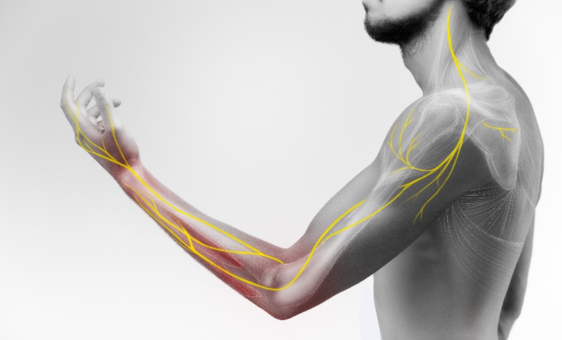
Peripheral neuropathy of the upper extremity, caused by trauma, entrapment, inflammation or systemic disease, can lead to significant disability. Diagnosis typically relies on clinical assessment and electrophysiological testing, but these approaches may not fully define lesion extent or precise anatomical location, especially in more complex presentations.
Imaging techniques such as MRN and high-resolution nerve ultrasound (HRUS) are increasingly used to address these gaps.
Diagnostic Overlap Remains Common
In a cohort of 800 patients with suspected upper extremity neuropathy, MRN and HRUS produced concordant findings in 59.9% of cases, indicating similar diagnostic information in the majority. However, discrepancies were observed in 40.1%, reflecting ongoing challenges in imaging interpretation.
After excluding non-relevant differences, 275 patients (34.4%) were identified where one modality provided additional clinically meaningful information. Within this subgroup, MRN accounted for 94.9% of cases with added value, while HRUS contributed in 5.1%.
Patterns Behind MRN’s Added Value
MRN was more likely to provide additional diagnostic insight in patients with proximal nerve lesions, where the odds were significantly higher compared with more distal disease. It also showed greater utility in cases involving multiple nerves or spanning more than one anatomical region.
Among these patients, clinical features were often complex: motor deficits were present in 77.0%, sensory symptoms in 55.6%, and more than half had multi-nerve involvement. Lesions frequently extended across multiple anatomical regions, further complicating localisation.
MRN’s ability to visualise deeper nerve structures and surrounding soft tissue may help explain its contribution in these cases.
HRUS Remains Valuable in Select Settings
Although less frequently associated with additional findings, HRUS retained clinical usefulness. Its added value was observed primarily in situations where MRN interpretation was limited by metal artefacts. Its accessibility and real-time imaging capabilities also support its continued role in evaluating more superficial nerve pathology.
Limitations Shape Interpretation
The findings should be interpreted cautiously for a few reasons. The study was conducted in a single tertiary centre and relied on clinician-assessed “perceived” diagnostic value, rather than an objective metric with external validation. Therefore, the study design limits conclusions about true diagnostic accuracy or cost-effectiveness.
Towards a Tailored Imaging Approach
These results support a more individualised use of imaging in upper extremity neuropathy. MRN may be considered in complex, proximal or multi-nerve cases, while HRUS remains a practical option when MRN is unavailable, contraindicated or affected by artefacts.
Reference
Brunnée M et al. High-resolution ultrasound vs. MR neurography in upper extremity neuropathies: exploratory analysis of perceived additional diagnostic value in routine clinical practice. Eur Radiol. 2026;DOI:10.1007/s00330-026-12545-0.
Featured image: BigBlueStudio on Adobe stock
- Author: