BACKGROUND AND AIMS
Fragility fractures represent a major global health burden in patients with osteoporosis, contributing to significant morbidity, mortality, reduced quality of life, and increased healthcare costs due to hospitalisation and surgical interventions.1-3 Although dual-energy X-ray absorptiometry (DXA) remains the current gold standard for assessing bone mineral density (BMD), it has important limitations. DXA primarily evaluates bone quantity and fails to capture early microarchitectural deterioration and qualitative changes in bone tissue, which are key determinants of bone strength.4,5 Consequently, a substantial proportion of fragility fractures occur in individuals classified as having osteopenia or even normal BMD, highlighting that DXA alone fails in predicting fracture risk.4 Current clinical guidelines emphasise the need for improved diagnostic strategies that integrate both bone density and clinical risk factors.2,6 This review aims to explore how advanced diagnostic tools can enhance fracture risk prediction and contribute to early preventive strategies, potentially reducing the need for surgical interventions.
MATERIALS AND METHODS
A narrative review of 65 recent studies was conducted to evaluate advancements in osteoporosis diagnostics and fracture risk prediction. The review focused on three principal domains: first, the limitations of conventional imaging modalities, such as DXA, in accurately predicting fracture risk;4-6 second, the clinical performance and potential advantages of emerging diagnostic techniques, particularly radiofrequency echographic multi-spectrometry (REMS), in assessing both BMD and bone quality;7-9 and third, strategies for early identification of patients at high risk for fragility fractures, including the integration of clinical risk factors and validated tools such as Fracture Risk Assessment Tool (FRAX®; University of Sheffield, UK).2,5 Relevant peer-reviewed articles were selected based on their methodological quality and clinical relevance.
RESULTS
The findings demonstrate that fragility fractures frequently occur in patients with osteopenia as assessed by DXA, underscoring the need for more sensitive and comprehensive methods for evaluating bone quality.4,5 REMS has emerged as a promising, non-ionising, portable and operator-independent diagnostic technique that evaluates both BMD and bone quality through the calculation of a fragility score. This method has demonstrated excellent precision with a root mean square coefficient of variation (RMS-CV) of less than 0.5%, enabling the detection of subtle changes in bone status over relatively short follow-up intervals.
Importantly, REMS allows for early identification of individuals at high fracture risk, facilitating timely implementation of preventive strategies such as pharmacologic therapy and fall prevention programmes, which are known to significantly reduce fracture incidence. Early intervention is crucial as it may decrease the occurrence of severe fractures that often require surgical management.While quantitative CT also provides valuable insights into trabecular bone structure and is less affected by degenerative changes or soft tissue artefacts, its use is limited by exposure to ionising radiation, higher cost, and reduced accessibility.10,11 In contrast, REMS offers a radiation-free alternative suitable for repeated measurements and bedside use, making it particularly advantageous in routine clinical practice and in vulnerable populations. These findings are summarised in Table 1, which compares DXA (with and without trabecular bone score [TBS]), quantitative CT, and REMS in terms of their ability to assess bone mineral density, bone quality, microarchitecture, precision, radiation exposure, and clinical applicability in osteoporosis management.
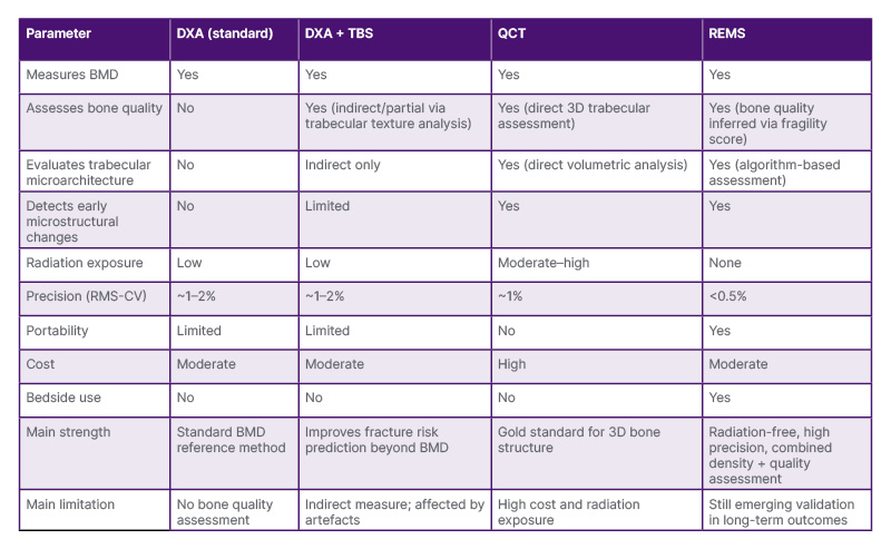
Table 1: Comparison of imaging modalities for assessment of BMD and bone quality in osteoporosis.
DXA: dual-energy X-ray absorptiometry; QCT: quantitative CT; REMS: radiofrequency echographic multi-
spectrometry; RMS-CV: root-mean-square coefficient of variation; TBS: trabecular bone score.
CONCLUSION
Advanced diagnostic tools such as REMS have the potential to overcome the limitations of conventional imaging techniques by providing a more comprehensive assessment of bone health, including both density and quality. By enabling earlier detection of bone deterioration and more accurate identification of high-risk individuals, these technologies support targeted preventive strategies that may significantly reduce fracture incidence and the subsequent need for surgical interventions. The integration of such innovative diagnostic approaches into clinical practice aligns with current international recommendations and may enhance fracture prevention and optimise patient care.