THE ASSESSMENT OF SPONDYLOARTHRITIS INTERNATIONAL SOCIETY (ASAS) and the SpA Research and Treatment Network (SPARTAN) have updated their key classification criteria for axial spondyloarthritis (axSpA).
Updated criteria and recommendations were presented at EULAR 2026, London, UK.
Current Classification Criteria
Currently used ASAS classification criteria for axSpA were developed in 2009.1
Despite their worldwide acceptance, they reportedly lack specificity.
Laure Gossec, President-Elect, EULAR, Kilchberg, Switzerland, told reporters today: “At the individual patient level, it was shown that these criteria were not very specific, meaning that you could actually reach the criteria but not have a diagnosis of spondyloarthritis.
“And, because low back pain – the key criterion for axial spondyloarthritis – is so frequent, concerning about 20% of the population in Europe, this low specificity led to overdiagnosis.”
ASAS and SPARTAN decided to update the 2009 criteria, based on a data-driven approach through the large international CLASSIC study.
Updated Classification Criteria
Overall, 1,015 patients referred to a rheumatologist with undiagnosed back pain indicative of axSpA were assessed and clinical, biological, and imaging criteria and their combinations were compared to the gold standard: the rheumatologist’s diagnosis for axSpA.
MRI of the sacroiliac joints, indicative of axSpA by assessment of both inflammatory and structural lesions, had the highest association with the diagnosis of axSpA.
HLA-B27, elevated CRP, and several clinical criteria including inflammatory back pain, inflammatory bowel disease, acute anterior uveitis, heel enthesitis, and psoriasis were also included in the revised criteria.
The final proposal achieved sensitivity of 79.5% and specificity of 90.4% in the validation dataset.
Presenting findings, Walter Maksymowych, University of Alberta, Edmonton, Canada, said: “The ASAS–SPARTAN revised classification criteria for axSpA emphasise the central role of imaging and incorporate a more focused set of clinical variables compared to the 2009 ASAS criteria.”
Updated Recommendations
It follows that, in a poster shared by Peter Mandl, Medical University of Vienna, Vienna, Austria, and colleagues at EULAR 2026, the original evidence-based EULAR recommendations on the use of imaging in the diagnosis and clinical management of both axial and peripheral SpA were updated.
Twelve key research questions on the role of imaging were adopted for the update.
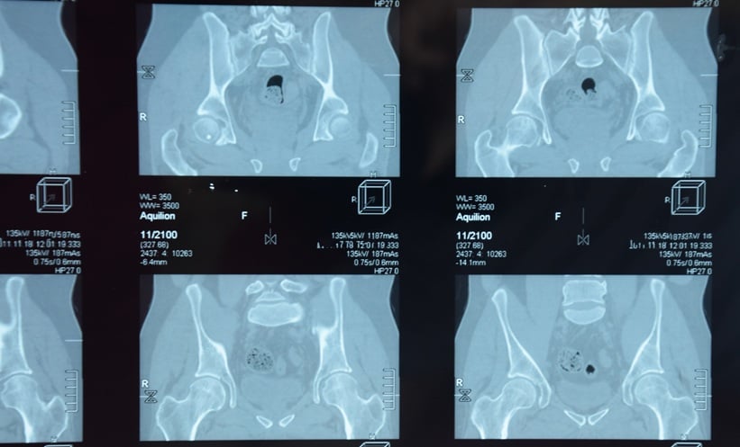
Imaging modalities included radiography, ultrasound, MRI, CT, PET, single photon emission computed tomography, dual-emission x-ray absorptiometry, and scintigraphy.
The task force formulated three new overarching principles, one new recommendation, and updates for nine of the originals.
The main changes included replacing radiography with MRI of the sacroiliac joint as the first imaging modality for diagnosing axSpA.
Radiography or preferably low-dose CT were not mentioned in the previous recommendations for diagnostic purposes but are now suggested as alternative modalities for the sacroiliac joint if MRI is not available or contraindicated.
For monitoring axSpA, MRI can be used to track sacroiliac and spinal inflammation and structural damage, while radiography is suitable for long term monitoring of structural damage in the spine.
For diagnosis of peripheral SpA, ultrasound or MRI remain the methods of choice.
In addition to inflammatory lesions, structural changes should now also be considered.
Ultrasound and MRI are also both now recommended for monitoring structural damage in peripheral SpA, along with radiography.
Reference
1 Rudwaleit M et al. The development of Assessment of SpondyloArthritis international Society classification criteria for axial spondyloarthritis (part II): validation and final selection. Ann Rheum Dis 2009;68(6):777–83.
Featured image: eleonimages on Adobe Stock
- Author: