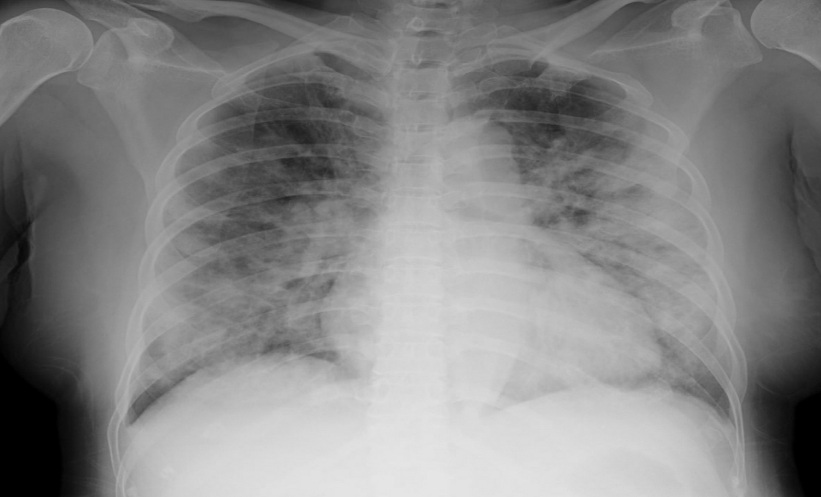
COVID-19 chest X-rays were more likely to show abnormalities in children nearly 10 months after SARS-CoV-2 infection than in uninfected peers, although low dose chest CT scans did not reveal significant differences between the groups, according to a longitudinal cohort study.
The findings suggest that any differences on imaging were confined to chest radiographs and were not linked to ongoing respiratory symptoms or asthma.
COVID-19 is caused by SARS-CoV-2 and primarily affects the respiratory system. While imaging findings during acute infection have been well characterised, the longer-term radiological effects in children following recovery remain less well understood.
Chest X-Rays Show Differences Nearly 10 Months After Infection
Researchers compared chest radiographs (CXR) and low-dose chest CT findings in 846 children (700 with laboratory-confirmed SARS-CoV-2 infection and 146 uninfected controls). Baseline imaging was performed a mean of 9.8 months after infection.
Three radiologists independently evaluated the images for recognised COVID-19-related abnormalities, including ground-glass opacities, reticular opacities, consolidation, nodules, perihilar thickening, pleural effusions and cystic changes.
Overall, radiological abnormalities were detected in 32.1% of infected children compared with 24.0% of controls. The difference was driven by CXR findings, while CT abnormalities did not differ significantly between the groups.
Children who had recovered from SARS-CoV-2 infection were more likely to have abnormalities on chest X-ray than uninfected participants (21.2% versus 12.4%; OR 2.44; 95% CI 1.19–5.02; p=0.016). This difference was primarily explained by a higher frequency of perihilar peribronchial thickening, which was observed in 17.4% of infected children compared with 10.1% of controls.
No Meaningful Differences in CT Findings and Symptoms
Low-dose chest CT identified abnormalities in 46.2% of infected participants and 42.1% of controls, with no statistically significant difference between the groups (OR 1.24; 95% CI 0.71–2.16; p=0.46).
The investigators also found that radiological abnormalities were not linked to pulmonary symptoms following recovery. Similarly, children with underlying asthma did not experience a higher frequency of imaging abnormalities than those without asthma.
The study found that children who had recovered from SARS-CoV-2 infection were more likely to have chest X-ray abnormalities nearly 10 months after infection, but these findings were limited to perihilar peribronchial thickening. No significant differences were observed on CT imaging, and radiological abnormalities were not linked to pulmonary symptoms following recovery or underlying asthma.
Reference
Zember J et al. Pediatric SARS-CoV-2 long term outcomes study: chest radiographic and computed tomography findings at baseline. Pediatr Radiol. 2026;DOI:10.1007/s00247-026-06701-3.
Featured image: Therads on Adobe Stock
- Author: