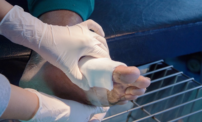
DIABETIC FOOT ULCER healing with negative pressure wound therapy (NPWT) may involve GRHL2 driven changes in cell fate, according to transcriptomic and single cell analyses that identified a potential molecular mechanism underlying wound repair.
Diabetic foot ulcers are a serious complication of diabetes associated with amputation, mortality, and substantial healthcare burden. Although NPWT is used to treat diabetic foot ulcers, the mechanisms through which it promotes wound healing remain incompletely understood.
Researchers analysed bulk RNA sequencing and single cell RNA sequencing data from diabetic foot ulcers. Differential expression analysis identified 975 dysregulated genes, of which 530 were upregulated and 445 were downregulated. Four machine learning algorithms were then applied to identify genes potentially activated by NPWT, with GRHL2 consistently selected across the analyses.
GRHL2 was primarily expressed in skin at both the RNA and protein levels. Single cell analysis of 13 diabetic foot ulcer samples further indicated that GRHL2 was predominantly expressed in keratinocytes.
NPWT Promoted Cell Conversion
Researchers assessed the mesenchymal epithelial transition capacity of keratinocytes and constructed differentiation trajectories using single cell analytical approaches.
The findings suggested that NPWT activated GRHL2, which subsequently drove the conversion of mesenchymal fibroblasts into supraspinous keratinocytes. Based on these results, the researchers established a model of mesenchymal fibroblast to supraspinous keratinocyte transition during diabetic foot ulcer repair.
Immunofluorescence analysis of clinical diabetic foot ulcer samples was subsequently used to validate GRHL2 expression. These findings were consistent with the transcriptomic analyses.
Diabetic Foot Ulcer Mechanism Offers New Insights
Together, the results led researchers to propose an NPWT, GRHL2, mesenchymal epithelial transition, and cell conversion signalling axis. This model suggested that the physical stimulation provided by NPWT could influence wound healing by regulating cell fate and conversion at the cellular level.
Molecular docking additionally identified parthenolide, MG 132, mitoxantrone, and irinotecan as compounds with potential activity relevant to diabetic foot ulcers. However, the abstract did not report experimental or clinical testing of these candidates, meaning their therapeutic efficacy remains to be established.
The findings provide a potential molecular explanation for how NPWT promotes diabetic foot ulcer healing and highlight GRHL2 mediated cell conversion as a mechanism for further investigation.
Reference
Wang S et al. Exploring the mechanism underlying negative pressure wound therapy in promoting the healing of diabetic foot ulcers via integrated multi-omics and bioinformatics approaches. Immunobiology. 2026;153228.
Image credit: Kirov1969 on Adobestock