AI in urological cancer diagnosis showed strong performance across CT, MRI, and ultrasound, with a systematic review and meta-analysis finding higher pooled diagnostic accuracy than the corresponding clinician estimates.
The analysis included 110 studies identified through searches of four electronic databases up to June 2026, reflecting the heavy reliance on imaging in urological cancer diagnosis and the growing interest in AI to improve radiological accuracy.
Does AI in Urological Cancer Diagnosis Matter for Clinical Practice?
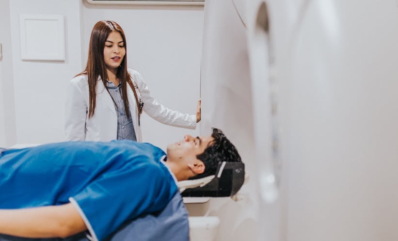
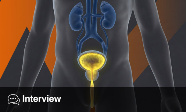
Urological cancers are cancers affecting the urinary system and male reproductive organs. Imaging with CT, MRI and ultrasound can support their detection and diagnosis, making reliable interpretation clinically important.
The researchers assessed AI algorithms and extracted clinician comparator data where available. They pooled sensitivity, specificity and area under the curve (AUC), a measure of overall diagnostic discrimination, using a bivariate random-effects model. Study quality was also assessed using the QUADAS-2 tool.
AI Showed Higher Specificity and AUC than Clinicians
AI models achieved pooled sensitivity of 0.85 and specificity of 0.83, with an AUC of 0.91. Clinicians had pooled sensitivity of 0.82, specificity of 0.68 and an AUC of 0.83.
The pooled estimates were therefore higher for AI than clinicians for specificity and AUC, while the difference in sensitivity was smaller. Subgroup analyses also indicated overall advantages for AI across cancer types and imaging modalities, although clinicians showed higher sensitivity in prostate cancer and MRI subgroups.
However, the clinician figures should not be interpreted as a definitive head-to-head comparison across all 110 studies. Comparator data were extracted when available, and the notes did not establish that AI and clinicians were assessed under identical conditions in every study. The findings therefore showed differences between pooled estimates rather than proving that AI was superior to clinicians in routine clinical practice.
What AI Could Mean for Urological Imaging
The findings supported AI as a potential adjunct to radiological workflows rather than a replacement for clinicians. They indicated that AI could potentially complement radiological assessment, although the analysis did not establish whether its use would improve patient outcomes or diagnostic pathways in practice. The researchers called for prospective, standardised, multi-centre evaluations to determine whether the observed diagnostic performance translated into clinical utility across diverse urological oncology tasks.
Reference
Shen Y et al. Artificial intelligence in the radiologic diagnosis of major urological cancers: a meta-analysis. World J Urol. 2026;DOI:10.1007/s00345-026-06635-3.
Featured image: magicmine on Adobe Stock