Abstract
Anomalous coronary arteries are uncommon but clinically important because certain patterns carry a higher risk of ischaemia and sudden death, whereas other variants, particularly anomalous left circumflex arteries (LCx) that run retroaortically, are usually benign, but can have important procedural and ischaemic implications. The authors present a 41-year-old man with tobacco exposure who presented with non-ST-elevation myocardial infarction whose catheter coronary angiogram demonstrated dual LCx: a native LCx arising from the left main, and a second accessory LCx arising near the right coronary ostium with a retroaortic course. This case highlights the diagnostic challenges associated with dual LCx anatomy, including recognition of an anomalous coronary origin during catheter angiography, and the complementary role of multimodality imaging. The clinical implications of this anatomy, including potential procedural considerations and management of associated coronary disease, are discussed in the context of the available literature.
Key Points
1. Dual, or ‘twin’, left circumflex arteries are exceptionally rare but clinically important coronary anomalies. This case highlights how failure to recognise an anomalous circumflex origin during coronary angiography may lead to incomplete anatomical assessment and underscores the value of multimodality imaging.2. Management of dual or anomalous circumflex anatomy should be tailored to patient stability, lesion accessibility, and ischaemic risk. While high-risk anatomy or culprit lesions may require percutaneous coronary intervention or surgery, many patients with distal or collateralised disease can be managed conservatively with optimised medical therapy and strict secondary prevention, including tobacco cessation.
3. Procedural awareness is essential: dual or anomalous circumflex arteries may complicate catheter selection and pose hazards during aortic and/or mitral valve surgery. Pre-procedural recognition and planning, supported by CT coronary angiography, can prevent catastrophic injury. The accompanying practical algorithm provides clinicians with stepwise guidance to integrate anatomy, imaging, and management into daily practice.
INTRODUCTION
Coronary artery anomalies are uncommon but clinically important because their anatomical variations may create diagnostic challenges and, in selected configurations, influence the risk of myocardial ischaemia or procedural complications. Dual, or ‘twin’, left circumflex arteries (LCx), in which separate circumflex vessels arise from different coronary origins, represent an exceptionally rare coronary anatomical variant. Recognition may be challenging during catheter coronary angiography, particularly when an anomalous vessel arises from the right coronary sinus or proximal right coronary artery and follows a retroaortic course.
The authors report a 41-year-old man who presented with non-ST-elevation myocardial infarction (NSTEMI) and was found to have dual LCx with distal occlusive disease and collateral filling. This case illustrates the importance of systematically evaluating the coronary anatomy when angiographic findings do not fully explain the clinical presentation and highlights the role of multimodality imaging and individualised management. Relevant literature is discussed to place the anatomical and clinical findings of this case in context and to highlight practical considerations for diagnosis and management.
CASE PRESENTATION
Patient Information and Findings
A 41-year-old man presented with a 4-month history of intermittent left precordial chest tightness associated with exertional light-headedness. The chest discomfort was non-radiating, ranged in severity from 3–7/10, lasted less than 20 minutes per episode, and was precipitated by exertion and relieved by rest. The frequency was intermittent and not specifically quantified. Over the preceding 4 months, the symptoms progressively worsened, prompting hospital evaluation. The exertional light-headedness was described as giddiness, lasted a few seconds, occurred exclusively with exertion, and resolved with rest. There was no history of syncope, palpitations, dyspnoea, diaphoresis, nausea, vomiting, or other associated symptoms. He was asymptomatic at the time of presentation.
The patient had previously been well, with no known history of hypertension, diabetes, dyslipidaemia, coronary artery disease, heart failure, arrhythmia, or other relevant cardiovascular or systemic comorbidity. He had never been hospitalised previously and had no history of surgery, invasive procedures, or significant trauma. He was not taking any regular prescription or over-the-counter medications before presentation. His family history was unremarkable for premature coronary artery disease, myocardial infarction, sudden cardiac death, cardiomyopathy, congenital heart disease, or other relevant cardiovascular disease.
The patient was a current cigarette smoker, smoking approximately 10 cigarettes per day for 20 years (10 pack-years), having started in his early twenties. He also reported infrequent smokeless tobacco use since his early twenties. He consumed alcohol occasionally, mainly beer, with hard liquor consumed only a few times per month. He denied recreational drug or other substance use. He predominantly followed a vegetarian diet, with occasional consumption of chicken and fish. He worked as a peasant farmer, primarily involved in livestock rearing and crop cultivation, representing a moderate level of habitual physical activity. There were no reported sleep-related symptoms.
On examination, he was comfortable, conscious, and oriented. His blood pressure was 110/70 mmHg, pulse rate 80 beats/min, and oxygen saturation 97% on room air; his BMI was 27.9 kg/m². He was neither pale nor cyanotic and had no jaundice, peripheral oedema, or clinical evidence of dehydration. The jugular venous pressure was not elevated, and peripheral pulses were normal and symmetrical. The apex beat was palpable in the fifth intercostal space at the mid-clavicular line and was not displaced. Heart sounds S1 and S2 were normal, with no S3 or S4, murmur, or pericardial rub. There were no clinical signs of heart failure or carotid bruits. Respiratory examination was normal, with no abnormal breath sounds. Abdominal examination was unremarkable, with no hepatomegaly or ascites. Neurological examination was normal, with no focal neurological deficit.
Investigations
Routine laboratory investigations were within normal limits. High-sensitivity troponin I was elevated at 1.76 ng/mL (reference <0.04 ng/mL) at presentation and increased to 1.89 ng/mL 6 hours later. The admission ECG showed sinus rhythm at 62 beats/min, with small Q waves in leads I and augmented vector left but no ST-segment or T-wave abnormalities. The PR interval was 180 ms, QRS duration 104 ms, and corrected QT interval 398 ms. Chest radiography was normal.
Transthoracic echocardiography demonstrated preserved left ventricular systolic function with a left ventricular ejection fraction of 55%. The left ventricular internal diameter in diastole was 47 mm and in systole was 33 mm. There were no regional wall motion abnormalities or significant valvular abnormalities. Right ventricular systolic function was preserved, with a tricuspid annular plane systolic excursion of 24 mm, and estimated pulmonary artery systolic pressure was 19 mmHg. There were no other structural cardiac abnormalities.
Coronary angiography demonstrated a normal left main coronary artery. The left anterior descending artery showed distal myocardial bridging. The native LCx originated from the left main coronary artery, while a second accessory LCx arose anomalously from the right coronary sinus near the ostium of the right coronary artery. The left coronary angiographic assessment in multiple projections demonstrated the native LCx and distal occlusive disease with collateral filling of the distal LCx territory. The accessory LCx was subsequently identified by right coronary injection, which demonstrated its anomalous origin near the right coronary ostium. The vessel was then selectively cannulated, and selective angiography demonstrated the accessory LCx and its distal occlusive disease with collateral filling. The anomalous coronary anatomy was established by catheter coronary angiography (Figure 1). CT coronary angiography was not performed.
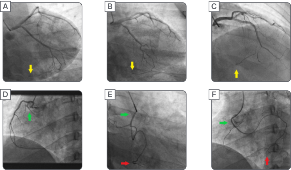
Figure 1: Coronary angiographic findings demonstrating dual left circumflex arteries.
A–C) Left coronary angiography showing the native left circumflex artery and collateral filling of the distal circumflex territory (yellow arrows); D) right coronary angiography demonstrating the accessory left circumflex artery arising near the ostium of the right coronary artery (green arrow); and E–F) selective angiography demonstrating distal occlusion of the accessory left circumflex artery (red arrows) with collateral filling from obtuse marginal branches.
Diagnosis
The diagnosis of NSTEMI was established on the basis of the patient’s typical exertional chest discomfort and elevated cardiac troponin concentrations in the absence of persistent ST-segment elevation. Coronary angiography demonstrated dual left circumflex arteries with distal occlusive disease and collateral filling of the distal circumflex territory. The accessory LCx arose from the right coronary sinus near the right coronary artery ostium and followed an anomalous course, while the native LCx originated from the left main coronary artery.
Management
Conservative management was selected because the occlusions involved distal branches of both LCx vessels, collateral circulation was established, left ventricular systolic function was preserved, and the patient remained haemodynamically stable without recurrent chest pain or clinical evidence of ongoing ischaemia or heart failure. He was treated with aspirin 75 mg once daily, clopidogrel 75 mg once daily, atorvastatin 40 mg once daily, nebivolol 2.5 mg once daily, and nicorandil 5 mg twice daily. Anticoagulation was not continued, and no angiotensin-converting enzyme inhibitor, angiotensin receptor blocker, angiotensin receptor-neprilysin inhibitor, or other additional cardiovascular medication was prescribed. Lifestyle modification was strongly reinforced, particularly complete cessation of cigarette smoking and smokeless tobacco, together with maintenance of regular physical activity and dietary risk factor modification.
Follow-Up and Outcome
At 1-year follow-up, the patient remained clinically well and reported no recurrence of chest pain, exertional dizziness, or other cardiovascular symptoms. There were no interim hospitalisations. He had successfully discontinued cigarette smoking and smokeless tobacco use. Repeat ECG showed no new ischaemic changes, and repeat transthoracic echocardiography continued to demonstrate preserved left ventricular systolic function. No repeat coronary angiography, CT coronary angiography, stress testing, or other new cardiac imaging was performed during follow-up.
Dual antiplatelet therapy was continued for 1 year, after which clopidogrel 75 mg once daily was continued as single antiplatelet therapy. At 1-year follow-up, his other medications were atorvastatin 20 mg once daily, nebivolol 2.5 mg once daily, and nicorandil 5 mg twice daily. The timeline of events is available as shown in Figure 2.
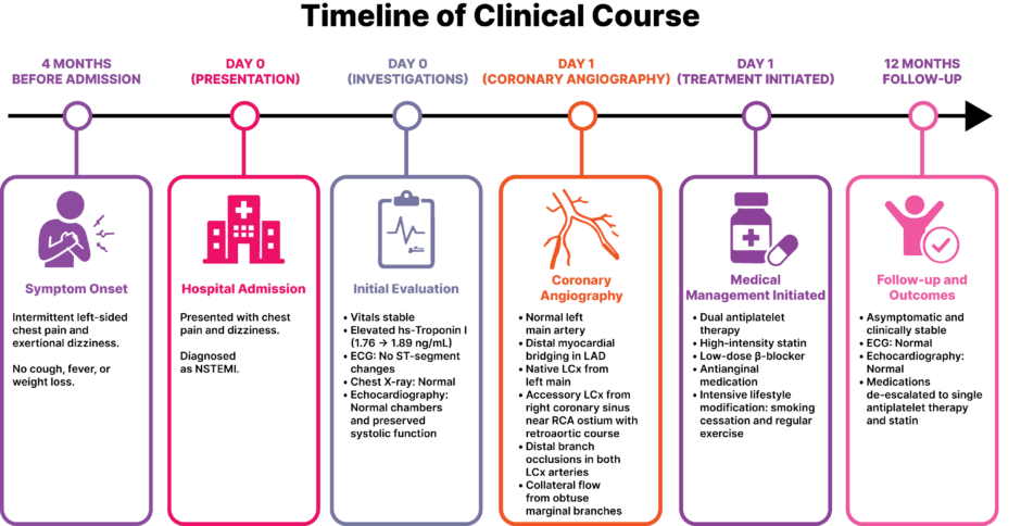
Figure 2: Timeline of events.
LAD: left anterior descending artery; LCx: left circumflex arteries; NSTEMI: non-ST-elevation myocardial infarction; RCA: right coronary artery.
DISCUSSION
This case illustrates a rare form of coronary artery anatomy in which two distinct left circumflex arteries arise from separate coronary origins and both demonstrate distal occlusive disease. The clinical importance of this finding extends beyond its rarity: failure to recognise an anomalous circumflex artery during coronary angiography may result in an incomplete anatomical assessment and may complicate interpretation of the patient’s clinical presentation.1 In this patient, identification of the accessory LCx arising near the right coronary ostium, together with collateral filling of the distal circumflex territory, was central to understanding the angiographic findings and supporting the decision for conservative management.
Dual LCx is exceptionally uncommon, with an incidence of approximately 0.19–0.29% reported in angiographic studies.2,3 Anomalous LCx arising from the right coronary sinus or proximal right coronary artery typically follows a retroaortic course.4 The available literature on dual LCx consists predominantly of individual case reports describing clinically significant presentations, including heart failure with stenotic twin LCx,5 acute myocardial infarction due to occlusion of a twin LCx,6 and acute coronary syndrome (ACS) associated with twin circumflex arteries.7 These reports provide useful context for the present case, but management should be guided primarily by the patient’s clinical status, the anatomy and location of coronary disease, and evidence of myocardial ischaemia rather than by the anatomical anomaly alone.8
Dual or anomalous LCx originating from the right coronary sinus or proximal right coronary artery typically pass posterior to the aortic root (retroaortic) to reach the lateral wall; this course is generally considered benign, as there is no interarterial compression between great vessels, but the anomalous ostium, acute angulation, and relationship to adjacent structures may influence atherosclerotic risk and procedural vulnerability.9 Embryologically, coronary artery anomalies are thought to reflect abnormal coronary budding and coronary sinus development; the clinical importance relates less to embryology and more to ostial morphology and early proximal course.10
Catheter coronary angiography remains the index test in ACS, but anomalous origins can be missed if the operator presumes normal left-system anatomy. If LCx perfusion is discordant with left injections (for example, ischaemia in the LCx territory but a normal left system injection), selective injections of the right coronary sinus or aortic root aortography should be performed to search for an accessory LCx or anomalous ostium. Failure to examine the right sinus systematically is the most common reason the accessory LCx is overlooked.11-13
CT coronary angiogram (CTCA) is now the reference standard for defining ostial origin, exact three-dimensional course, and relationship to neighbouring structures, information that often cannot be fully captured by two-plane angiography. CTCA is therefore recommended when catheter coronary angiography is ambiguous or when the precise anatomical course will change management, for example, in preoperative planning. CTCA is also superior for preoperative planning prior to valve or aortic surgery.4,14
A practical bedside clue to a retroaortic LCx is the retroaortic anomalous coronary sign on transthoracic echocardiography, an echogenic tubular structure posterior to the aortic root seen in specific views. Several observational reports confirm its utility as a screening sign that should prompt CTCA confirmation. Awareness of this sign can accelerate diagnosis when CTCA access is not immediately available.15,16
Most retroaortic LCx anomalies are clinically silent, yet the accessory LCx can develop atherosclerotic lesions and present as ACS; case literature documents both conservative and interventional outcomes. The decision to revascularise should therefore be based primarily on the clinical presentation, lesion severity and accessibility, evidence of ischaemia, haemodynamic status, and the anatomical risk profile rather than on the presence of a coronary anomaly alone.8,17 High-risk anatomies (interarterial/intramural, slit-like ostium) are associated with sudden cardiac death and warrant more aggressive interventions; retroaortic LCx is typically not in that high-risk group but holds procedural relevance.9,18
This case included distal left anterior descending artery myocardial bridging, a commonly encountered variant that occasionally associates with ischaemia, especially when deep and/or long bridges coexist with atherosclerosis. Functional assessment by stress testing and/or invasive physiology may help tailor therapy when myocardial bridging is suspected as a contributor to symptoms.19,20
For patients with anomalous LCx anatomy presenting with ACS, initial management should follow established ACS principles rather than being determined by the coronary anomaly alone. The 2023 European Society of Cardiology (ESC) Guidelines for the management of ACS recommend early clinical risk stratification, antithrombotic therapy, and an invasive strategy according to the patient’s ischaemic risk and clinical status.21 The 2025 American College of Cardiology (ACC)/American Heart Association (AHA)/American College of Emergency Physicians/National Association of EMS Physicians/Society for Cardiovascular Angiography and Interventions (SCAI) Guideline similarly recommends guideline-directed antithrombotic therapy, risk-based invasive management, and secondary prevention for patients with ACS, including NSTEMI.22 In patients with anomalous coronary anatomy, the 2018 AHA/ACC Adult Congenital Heart Disease Guideline emphasises assessment of the anomalous coronary origin, ostial and proximal anatomy, symptoms, and evidence of ischaemia when determining management; however, these recommendations primarily address anomalous aortic origin of a coronary artery and do not specifically address dual or twin LCx.17 Thus, management of dual LCx presenting with ACS requires extrapolation from general ACS and anomalous-coronary principles, supplemented by the limited case-based literature.
When a culprit lesion in an anomalous LCx is technically accessible and clinically significant, PCI may be considered, although anomalous ostial anatomy and catheter engagement may increase procedural complexity.18,23 Conversely, in a haemodynamically stable patient with distal, collateralised disease, preserved ventricular function, and no evidence of ongoing ischaemia, a conservative strategy with guideline-directed medical therapy and secondary prevention may be reasonable.8,23
Recognition of dual or anomalous LCx anatomy during catheter coronary angiography requires a systematic approach. When ischaemia in the LCx territory is suspected but left-system angiography appears normal, selective right sinus injections or even aortic root angiography should be performed to identify a possible accessory LCx arising near the right coronary artery.24 Engaging the ostium can be technically challenging because of its acute angulation and proximity to the right coronary artery, often necessitating alternative catheter shapes and gentle manipulation to avoid vessel trauma; preprocedural CTCA is particularly useful in planning such interventions and anticipating support limitations.18 For surgeons, preoperative recognition of a retroaortic LCx is crucial, as this vessel can be injured during valvular procedures. CTCA should therefore be performed whenever an anomaly is suspected preoperatively, enabling modification of surgical techniques such as altered annuloplasty suture placement or root remodelling to prevent injury.25,26 Together, these interventional and surgical precautions underscore the importance of multimodality imaging and proactive planning when dealing with anomalous circumflex anatomy.
In this patient, the distal location of the occlusions, established collateral circulation, preserved LV systolic function, haemodynamic stability, and absence of recurrent ischaemia supported a conservative strategy. This approach is consistent with the broader principle in both ESC and ACC/AHA ACS guidance that invasive management should be individualised according to clinical and ischaemic risk, while revascularisation decisions should also account for lesion accessibility and the expected benefit of intervention.21-23 However, because neither guideline specifically addresses dual LCx, the decision in this case was necessarily individualised using these general principles together with the limited published experience of twin LCx.27,28
A stepwise algorithm for evaluating suspected dual or anomalous LCx is shown in Figure 3. When ischaemia is suspected in the circumflex territory but left coronary angiography appears normal, selective right sinus or root injections should be performed. If an accessory LCx is identified, documentation of its ostial origin and proximal course is essential. Risk stratification then guides management. Patients with interarterial or intramural courses, or those with ongoing ischaemia or high GRACE scores, should be considered for early revascularisation with percutaneous coronary intervention or surgery. Conversely, patients with distal, collateralised disease who remain haemodynamically stable are usually best managed conservatively with optimised medical therapy. If diagnostic uncertainty persists, targeted non-invasive ischaemia testing or CTCA can refine the assessment. Ultimately, treatment decisions should integrate anatomic findings, ischaemic risk, and overall patient stability to achieve the safest and most effective outcome.
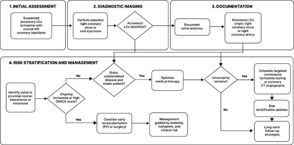
Figure 3: Practical approach to recognition, documentation, risk stratification, and management of suspected anomalous circumflex coronary arteries.
GRACE: Global Registry of Acute Coronary Events; LCx: left circumflex artery; PCI: percutaneous coronary interventions.
CONCLUSION
Dual or ‘twin’ LCx are exceedingly rare and usually benign, yet they can occasionally present with ACS. Recognition requires a high index of suspicion, particularly when LCx territory ischaemia is evident but left-system angiography appears normal; in such situations, careful interrogation of the right coronary sinus or aortic root is warranted to avoid overlooking an accessory vessel. Modern imaging plays an essential role: CTCA reliably defines the anomalous ostium and confirms the retroaortic course, while transthoracic echocardiography may reveal the retroaortic anomalous coronary sign as a bedside clue. Management should be individualised according to the patient’s clinical and ischaemic risk, the location and accessibility of coronary disease, and the anatomical characteristics of the anomalous vessel. General ACS management should follow contemporary ESC and ACC/AHA recommendations, while decisions specific to anomalous coronary anatomy should be informed by established anomalous-coronary guidance and the limited evidence available for dual LCx. Ultimately, this case underscores the importance of prompt recognition, accurate anatomical delineation, and individualised treatment of dual LCx anatomy, together with rigorous secondary prevention including tobacco cessation.