NEUROIMAGING highlights a range of neurological findings after lightning injury (LI), including brain edema, intracerebral or subarachnoid bleeding, and ischemia. In the retrospective study, results also revealed that white matter lesions were common, although their significance in relation to LI remains unknown.
Lightning Injuries
Lightning injuries can affect multiple organs. These include the peripheral nervous system and central nervous system (CNS). However, there is a scarcity of neuroimaging studies in LI, and it remains unclear whether lightning injury causes additional changes in the CNS.
Researchers carried out a retrospective review to assess patients who were treated in two tertiary neurology referral centres between 2004–2024. Patients referred to these units were treated for sequelae of LI and underwent MRI or computed tomography of the CNS. Additionally, the authors conducted a narrative literature search in order to identify other studies looking at neuroimaging after LI.
Neuroimaging Findings
In total, 31 patients were assessed. The median age was 38 years, and 39% of the patients were female. Thirty patients underwent cranial neuroimaging, and 14 underwent spinal MRI. The median delay between LI and the first neuroimaging assessment was 67 days.
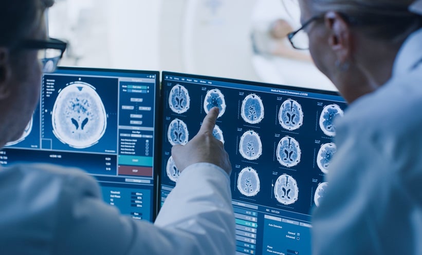
Patients who underwent cranial MRI or CT showed a range of neuroimaging findings. Two patients had brain edema, one of whom also had subarachnoid bleeding. One patient had laminar cortical necrosis after resuscitation, while another had hyperintensities in the hippocampi. Cranial MRI showed nonspecific white matter lesions in 14 patients. Additionally, of the 14 patients who underwent spinal MRI, six had intramedullary spinal cord lesions.
In a review of 54 published patients, pathological neuroimaging findings were identified in 44% of cases. Researchers reported that the main findings were hypoxic encephalopathy following cardiac arrest, intracerebral bleeding, cerebral ischemia, brain edema, and subarachnoid bleeding.
Conclusions
Overall, typical neuroimaging findings following LI included brain edema, intracerebral or subarachnoid bleeding, ischemia, hypoxic encephalopathy, and intramedullary spinal cord lesions.
The researchers also noted that white matter lesions were common; however, the significance of these lesions remains unknown. This is due to the rarity of clinical correlates and the inability to rule out pre-existing pathologies.
Reference
Steinberg T et al. Neurological and neuroimaging sequelae of lightning injury: a retrospective study and literature review. Neurol Res Pract. 2026;8:77.
Featured Image: FotoBastek on Adobe Stock