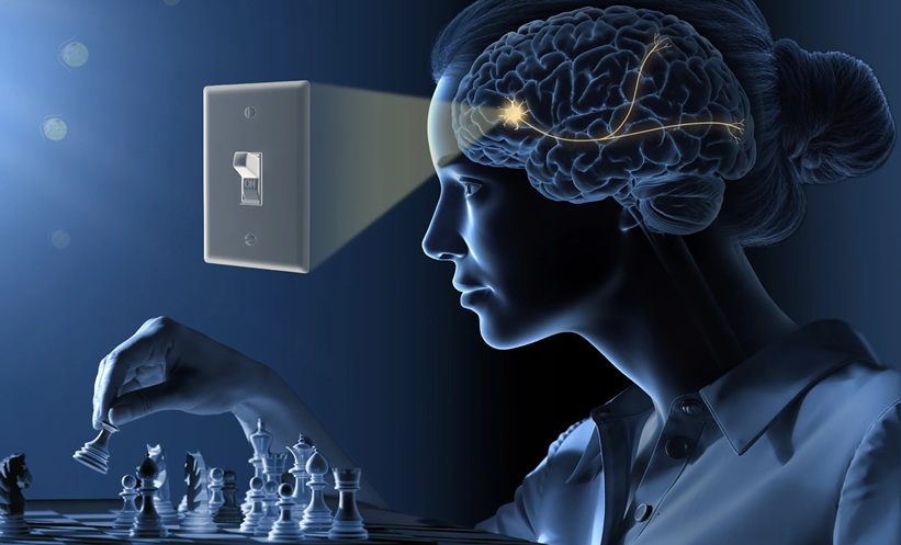
THE NOBEL PRIZE in Physiology or Medicine 2026 has been awarded to a team of scientists behind optogenetics, a breakthrough method that makes it possible to show how nerve cells in the brain shape memories, feelings and behaviour.
Announced today, Karl Deisseroth, Peter Hegemann, Georg Nagel, who have been recognised for their discovery of a molecular switch for nerve cells in the living brain, are credited with laying the foundation of a new era in neuroscience.
Using light, researchers can now switch individual neural circuits on or off in nerve cells, a technique that is fundamentally transforming our understanding of the brain.
Ligh Reactive Algae Offered Key to Extraordinary Protein
The era-defining discovery began when Peter Hegemann, Humboldt University of Berlin, Germany, began investigating how Chlamydomonas, a single-celled alga, is able to recognise and swim towards a light source.
Using minuscule electrodes, he measured electrical signals that arose in the Chlamydomonas when it’s eye spot – or sensor for light – was illuminated, finding an electrical impulse occurred just half a millisecond after this moment.
This process is more than 20 times faster than the complex chemical chain reaction that is triggered when light reaches the human eye. The final protein in this long chain forms an opening in the cell surface through which ions can flow, known as an ion channel.
Hegemann theorised that a single protein complex within the Chlamydomonas both captured light and acted as an ion channel, despite scepticism from other members of the research community.
Later, working alongside colleague George Nagel, University of Würzburg, Germany in 2000, using egg cells from frogs to study proteins, the pair discovered the extraordinary protein present in the single-celled algae, known as channelrhodopsin-2.
Nagel’s characterisation of channelrhodopsin-2 showed that it reacted exceptionally strongly and rapidly to a pulse of light.
When copied into batches of frog eggs, a channel opened through the surface of the egg within 0.2 milliseconds when exposed to light, through which positively charged ions flowed creating an electrical signal.
They were able to recreate this effect after introducing the gene for channelrhodopsin-2 into embryonic human kidney cells and in kidney cells from hamsters.
The Birth of Optogenetics
Neuroscientist Karl Deisseroth, The Howard Hughes Medical Institute and Stanford University, USA, was inspired to study how nerve cells function in a living brain in order to better understand psychiatric diseases he encountered working at a clinic.
He gained access to the to the DNA encoding the channelrhodopsin-2 protein through Nagel and Heggeman, eventually, introducing it into rat nerve cells cultured in petri dishes. When exposed to blue light, the nerve reacted immediately.
The light provoked a nerve signal that could be propagated to other nerve cells. Using this technique, Deisseroth successfully activated nerve cells in the brains of living mice – a turning point in neuroscience.
He introduced the channelrhodopsin-2 into the nerve cells of the motor cortex of mice. By illuminating these nerves cells with an optic fibre fed into the mouse’s brain, they were able to control movements of the mouse whiskers. They later used the same technique to awake sleeping mice.
This method for controlling nerve signals with light became known as optogenetics, and it has rapidly gained global impact.
Using optogenetics, researchers have been able to reveal neural circuits governing specific memories, feelings, and behaviours relevant for neurological and psychiatric disorders.
In clinical medicine, researchers are using the method in attempts to restore sight in people with visual impairment.
Overall, the trio are credited with fundamentally altering our understanding of the brain, according to the Nobel Prize, transforming neuroscience and helping to unravel one of humanity’s greatest mysteries: the human brain.