Abstract
Background: Primary endometrial signet ring cell adenocarcinoma is an exceptionally rare histological variant of endometrial carcinoma, with only a limited number of cases reported to date. Because signet ring cell morphology is more commonly associated with metastatic carcinomas originating from the gastrointestinal tract or breast, establishing a primary endometrial origin represents a significant diagnostic challenge requiring comprehensive clinicopathological and immunohistochemical evaluation.
Case Presentation: A 67-year-old multiparous postmenopausal woman presented with dysuria and urinary frequency without vaginal bleeding. Transvaginal ultrasonography demonstrated a 7 mm irregular endometrium. Endometrial Pipelle biopsy revealed poorly cohesive atypical epithelioid cells with focal signet ring cell morphology. Serum tumour markers were within normal limits. Extensive diagnostic investigations, including gastroscopy, colonoscopy, breast imaging, pelvic MRI, and PET-CT, excluded an extragenital primary malignancy. The patient subsequently underwent total laparoscopic hysterectomy with bilateral salpingo-oophorectomy. Histopathological examination of the surgical specimen revealed no residual carcinoma. Immunohistochemical analysis demonstrated diffuse pancytokeratin (PCK) positivity, while PAX8, GATA3, p63, and human placental lactogen were negative, supporting the final diagnosis after exclusion of metastatic disease. The patient remains disease-free 23 months after surgery.
Conclusion: Primary endometrial signet ring cell adenocarcinoma should be considered in the differential diagnosis of uterine tumours exhibiting signet ring morphology. Careful exclusion of metastatic disease through comprehensive clinical evaluation, imaging studies, and immunohistochemical analysis is essential for accurate diagnosis and appropriate management. This case contributes additional evidence regarding the clinicopathological characteristics and favourable short-term outcome of this exceptionally rare malignancy.
Key Points
1.Primary endometrial signet ring cell adenocarcinoma is an exceptionally rare malignancy. Because signet ring cell morphology is far more commonly associated with metastatic gastrointestinal or breast carcinoma, establishing the correct primary origin requires meticulous clinicopathological correlation and comprehensive immunohistochemical evaluation.2.This report describes a 67-year-old postmenopausal woman with an incidental diagnosis of primary endometrial signet ring cell adenocarcinoma. Extensive imaging, endoscopic examinations, and immunohistochemical analyses successfully excluded an extragenital primary tumour, allowing an accurate diagnosis of this rare entity.
3.Clinicians should always exclude metastatic disease before diagnosing primary endometrial signet ring cell carcinoma. A multidisciplinary diagnostic approach integrating imaging, pathology, and immunohistochemistry is essential for appropriate treatment planning and favourable clinical outcomes.
INTRODUCTION
In developed countries, endometrial adenocarcinoma is one of the most common malignancies of the female genital tract.1 It is typically diagnosed in postmenopausal women following endometrial sampling performed for abnormal uterine bleeding, although asymptomatic cases may occasionally be encountered. Several uncommon histological variants have been described, including villoglandular, secretory, ciliated cell, and tumours with squamous differentiation.2 Among these rare variants, primary endometrial signet ring cell adenocarcinoma represents an exceptionally uncommon histopathological subtype. According to the availableliterature, the first case was reported by Mooney et al.3 in 1997.
Signet ring cell morphology within the endometrium presents a significant diagnostic challenge because it is more commonly associated with metastatic carcinomas originating from the gastrointestinal tract or breast than with a primary endometrial malignancy. Therefore, establishing the diagnosis requires a comprehensive clinicopathological evaluation, including radiological imaging, endoscopic investigations, and immunohistochemical analysis to exclude an extragenital primary tumour.
To provide an updated overview of this exceptionally rare entity, the authors performed a comprehensive literature search on PubMed and Google Scholar with the keywords ‘Primary Endometrial Carcinoma with Signet Ring Cells’. All available reports were reviewed without language or time restrictions. The clinical characteristics of the previously published cases are summarised in Table 1.
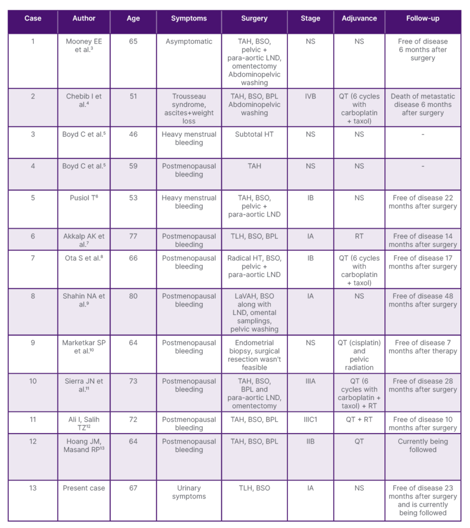
Table 1: Reported cases of primary endometrial signet ring cell adenocarcinoma.
BPL: bilateral pelvic lymphadenectomy; BSO: bilateral salpingo-oophorectomy; HT: hysterectomy; LaVAH: laparoscopically assisted vaginal hysterectomy; LND: lymph node dissection; NS: not specified; QT: chemotherapy; RT: radiation therapy; TAH: total abdominal hysterectomy; TLH: total laparoscopic hysterectomy.
Herein, the authors present the case of a 67-year-old postmenopausal woman diagnosed with primary endometrial signet ring cell adenocarcinoma and discuss the clinicopathological findings in the context of the existing literature, with particular emphasis on the diagnostic role of immunohistochemistry in distinguishing primary endometrial disease from metastatic carcinoma.
PATIENT INFORMATION
A 67-year-old multiparous woman (gravida 11, para 11; all spontaneous vaginal deliveries) presented to the gynaecology outpatient clinic with complaints of dysuria and increased urinary frequency. She had been postmenopausal for 7 years and denied postmenopausal vaginal bleeding or other gynaecological symptoms.
Her medical history was significant for Type 2 diabetes, which was managed with oral antidiabetic medication. She had no history of previous surgical procedures. Routine cervical cancer screening was unremarkable, with a negative human papillomavirus test performed 3 years before presentation and a negative Papanicolaou smear at the age of 66.
The patient had no personal or family history of gynaecologic, gastrointestinal, or breast malignancy. She was a non-smoker and reported no alcohol consumption. Her BMI was 33.27 kg/m², consistent with obesity. No other relevant psychosocial or genetic history was identified.
CLINICAL FINDINGS
The patient was in good general condition, and her vital signs were stable at presentation. Abdominal examination was unremarkable.
On gynaecological examination, the vulva, vagina, and cervix appeared normal on speculum examination, with no visible lesions or abnormal discharge. Bimanual pelvic examination revealed a freely mobile, anteverted uterus of postmenopausal size without adnexal masses or parametrial involvement. Both adnexa were clinically unremarkable.
Transvaginal ultrasonography demonstrated an anteverted uterus with an irregular endometrium measuring 7 mm in thickness. Both ovaries appeared atrophic, and no adnexal pathology or free pelvic fluid was identified.
TIMELINE
The timeline of the patient’s clinical course, including diagnostic evaluation, treatment, and follow-up, is summarised in Table 2.
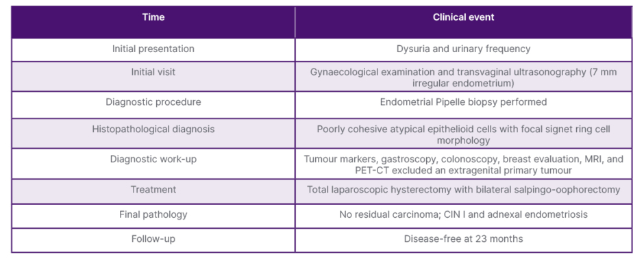
Table 2: Timeline of the patient’s clinical course, diagnostic evaluation, treatment, and follow-up.
CIN: cervical intraepithelial neoplasia.
DIAGNOSTIC ASSESSMENT
Endometrial sampling was performed using a Pipelle biopsy after transvaginal ultrasonography demonstrated an irregular endometrium measuring 7 mm in thickness. Histopathological examination revealed poorly cohesive atypical epithelioid cell clusters with focal signet ring cell morphology embedded within a fibrinous background, raising suspicion for signet ring cell carcinoma. Immunohistochemical analysis demonstrated diffuse pancytokeratin (PCK) positivity, confirming the epithelial nature of the tumour cells. The neoplastic cells were negative for PAX8, GATA3, p63, and human placental lactogen, with a Ki-67 proliferation index of approximately 60–70% (Figure 1).
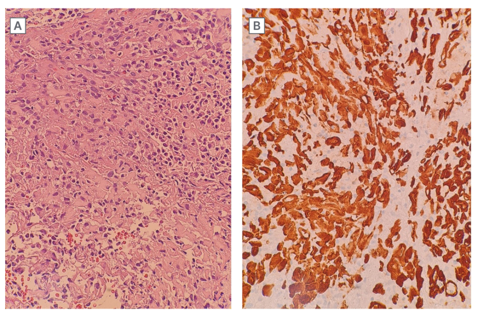
Figure 1: Side-by-side microscopic images at ×40 magnification.
A) Haematoxylin and eosin-stained section showing clusters of atypical epithelioid cells with intracytoplasmic mucin, consistent with signet ring cell morphology, scattered within a fibrinous background. B) Immunohistochemical staining for PCK demonstrating diffuse and strong cytoplasmic positivity in tumour cells, confirming the epithelial nature of the tumour cells.
PCK: pancytokeratin.
Because signet ring cell morphology is far more commonly associated with metastatic carcinoma than with a primary endometrial neoplasm, an extensive diagnostic work-up was performed. Serum carcinoembryonic antigen (CEA), cancer antigen-125 (CA-125), cancer antigen 15-3 (CA15-3), and cancer antigen 19-9 (CA19-9) levels were all within normal limits. Gastroscopy, colonoscopy, breast examination, mammography, pelvic MRI, and PET-CT revealed no evidence of a gastrointestinal, breast, or other extragenital primary malignancy.
The principal diagnostic challenge was distinguishing a primary endometrial tumour from metastatic signet ring cell carcinoma because of the exceptional rarity of this histological subtype and its nonspecific clinical presentation. Integration of the histopathological findings with immunohistochemistry, endoscopic evaluation, and comprehensive radiological assessment supported the diagnosis of primary endometrial signet ring cell adenocarcinoma. Based on the overall clinicopathological findings, the disease was classified as International Federation of Gynecology and Obstetrics (FIGO) Stage IA.
THERAPEUTIC INTERVENTION
Following multidisciplinary evaluation and exclusion of an extragenital primary malignancy, surgical management was planned after detailed patient counselling and written informed consent. The patient underwent total laparoscopic hysterectomy with bilateral salpingo-oophorectomy. Intraoperatively, the uterus was of postmenopausal size, and both ovaries appeared atrophic. Endometriotic foci were observed on the serosal surface of the uterus and both adnexa. No gross evidence of extrauterine tumour dissemination, peritoneal implants, or enlarged pelvic lymph nodes was identified. Macroscopic examination of the hysterectomy specimen revealed no gross evidence of malignancy (Figure 2).
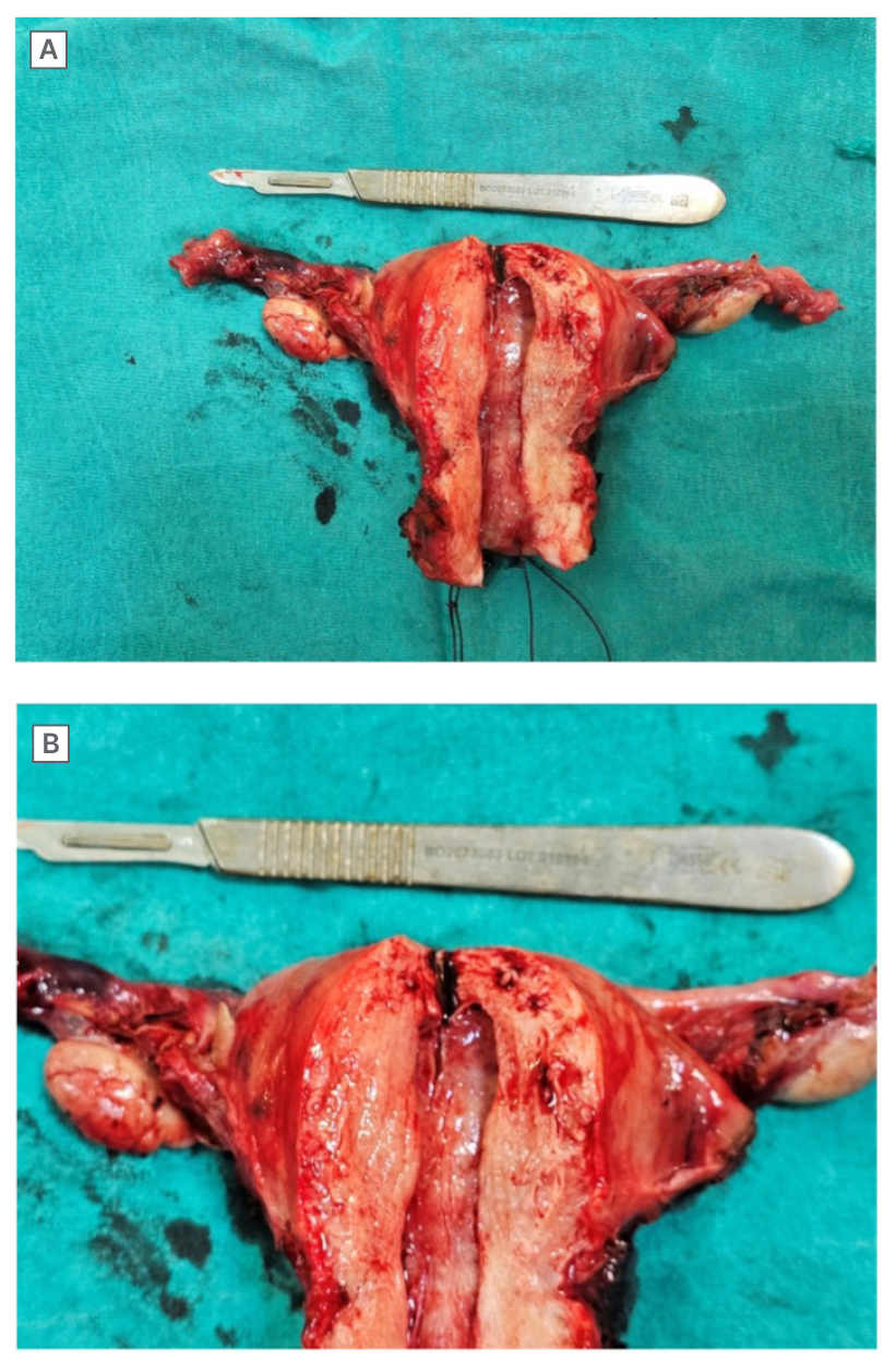
Figure 2: Gross appearance of the hysterectomy specimen.
A) Wider view of the hysterectomy specimen. B) Closer view of the opened uterine specimen and endometrial cavity. No gross tumour mass or suspicious lesion was identified.
Histopathological examination of the hysterectomy specimen demonstrated no residual carcinoma. Incidental findings included cervical intraepithelial neoplasia Grade I and adnexal endometriosis. In view of the absence of residual disease and the comprehensive exclusion of an extragenital primary malignancy, no additional surgical procedure or adjuvant oncological treatment was recommended.
FOLLOW-UP AND OUTCOMES
The postoperative course was uneventful, and the patient was discharged in good general condition. No perioperative or postoperative complications occurred.
During follow-up, no adjuvant chemotherapy or radiotherapy was administered because the hysterectomy specimen showed no residual carcinoma and there was no evidence of metastatic disease. Clinical follow-up consisted of regular clinical examinations with imaging performed when clinically indicated.
At 23 months after surgery, the patient had no evidence of local recurrence or distant metastasis, and her overall clinical condition was satisfactory.
DISCUSSION
Primary endometrial signet ring cell adenocarcinoma is an exceptionally rare histological subtype of endometrial carcinoma. Because signet ring cell morphology is far more frequently encountered in metastatic carcinomas of the gastrointestinal tract and breast than in primary uterine neoplasms, its identification within the endometrium should initially raise suspicion for metastatic disease rather than a primary malignancy.7 Consequently, establishing the diagnosis requires meticulous clinicopathological correlation supported by comprehensive radiological, endoscopic, and immunohistochemical evaluation.
The greatest diagnostic challenge in the present case was differentiating a primary endometrial tumour from metastatic signet ring cell carcinoma. Histopathological identification of signet ring cells alone is insufficient to determine the site of origin because morphologically identical tumours may arise from the stomach, colorectum, breast, urinary bladder, or other organs.7,11 Therefore, the patient underwent extensive investigations, including gastroscopy, colonoscopy, breast examination, mammography, pelvic MRI, and PET-CT, all of which failed to identify an extragenital primary malignancy. This comprehensive evaluation strongly supported the diagnosis of a primary endometrial carcinoma after exclusion of an extragenital primary malignancy.
Immunohistochemistry is an important adjunct in the differential diagnosis of signet ring cell tumours involving the uterus.7,11 GATA3 is widely recognised as a sensitive marker of breast carcinoma and is useful in excluding metastatic mammary tumours.9,10,14 Likewise, PAX8 has been reported as a valuable Müllerian marker supporting a primary gynaecologic origin.10,15 However, PAX8 was negative in this patient. Immunohistochemical analysis demonstrated diffuse PCK positivity, confirming the epithelial nature of the tumour, whereas GATA3, p63, and human placental lactogen were negative. Therefore, the diagnosis was established through integration of the histopathological findings, immunohistochemical profile, and comprehensive exclusion of an extragenital primary malignancy rather than on a single immunohistochemical marker.
The terminology of signet ring cell tumours arising in the endometrium remains controversial. Pusiol6 proposed that tumours containing conventional endometrioid adenocarcinoma with only focal signet ring cell differentiation should be designated endometrial adenocarcinoma with signet ring cells, whereas the term primary signet ring cell adenocarcinoma should be reserved for tumours composed almost entirely of signet ring cells.6 In this case, the biopsy specimen demonstrated poorly cohesive atypical epithelioid cells with focal signet ring cell morphology, emphasising the diagnostic complexity of this rare entity.
Among reported cases, most patients were postmenopausal women presenting with abnormal uterine bleeding.3-5,7-13 In contrast, this patient presented only with dysuria and urinary frequency, without postmenopausal bleeding, leading to an incidental diagnosis following evaluation of a mildly irregular endometrium. This uncommon clinical presentation suggests that primary endometrial signet ring cell adenocarcinoma may remain clinically silent and should also be considered in the differential diagnosis of asymptomatic postmenopausal women with suspicious endometrial findings.
Another noteworthy aspect of the present case was the complete absence of residual carcinoma in the hysterectomy specimen despite the initial biopsy confirming signet ring cell adenocarcinoma. One possible explanation is complete excision of a very small lesion during Pipelle sampling; however, spontaneous absence of residual tumour cannot be confirmed with certainty.
Because of its rarity, no standardised treatment strategy has been established for primary endometrial signet ring cell adenocarcinoma. Most reported patients have undergone hysterectomy with bilateral salpingo-oophorectomy, frequently combined with pelvic and/or para-aortic lymphadenectomy.3,4,7,8,11-13 Adjuvant chemotherapy, radiotherapy, or combined chemoradiotherapy has been administered inconsistently, generally according to disease stage and institutional preference.4,7,8,10-13 In this patient, comprehensive preoperative evaluation demonstrated no evidence of metastatic disease, and the hysterectomy specimen showed no residual carcinoma. Therefore, neither additional surgical staging nor adjuvant oncologic treatment was considered necessary.
The principal strength of this report is the comprehensive multidisciplinary diagnostic evaluation performed before establishing the diagnosis of primary endometrial signet ring cell adenocarcinoma. Furthermore, the authors provide an updated review of all previously published cases and summarise the clinicopathological characteristics. The main limitation is the inherent nature of a single case report, which precludes definitive conclusions regarding prognosis or optimal management. Nevertheless, given the extreme rarity of this entity, each additional well-documented case contributes valuable evidence that may improve future diagnostic accuracy and therapeutic decision-making.
CONCLUSION
Primary endometrial signet ring cell adenocarcinoma is an exceptionally rare malignancy that should only be diagnosed after meticulous exclusion of metastatic disease. A multidisciplinary approach integrating histopathology, immunohistochemistry, endoscopic evaluation, and comprehensive imaging is essential for accurate diagnosis and appropriate management. Further accumulation of well-documented cases is needed to better define the biological behaviour, prognosis, and optimal management of this exceptionally rare malignancy.
PATIENT PERSPECTIVE
The patient expressed satisfaction with the diagnostic process and surgical treatment. She reported relief after learning that no residual tumour was identified and has remained satisfied with her recovery during follow-up visits.