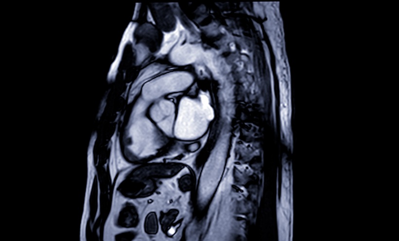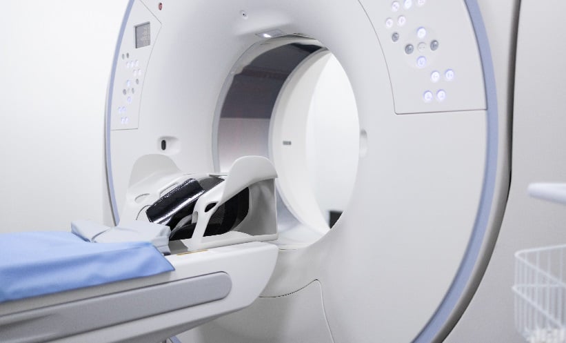
INVASIVE lobular breast cancer (ILC) remains a diagnostic challenge, particularly in assessing the spread of disease to axillary lymph nodes (ALNs). Accurate staging is critical for treatment planning, but conventional imaging often struggles to detect metastasis reliably in this subtype. This prospective study investigated the utility of a specialised imaging technique, 18F-fluoroestradiol (18F-FES) PET/CT, in identifying ALN metastasis in patients newly diagnosed with oestrogen receptor-positive ILC. Notably, the scan demonstrated 100% specificity when read visually and semiquantitatively.
Between August 2023 and August 2024, 20 women with newly diagnosed oestrogen receptor-positive ILC and suspected or confirmed ALN involvement were enrolled in the feasibility study. All participants were scheduled for axillary surgery. 18F-FES PET/CT scans were interpreted both visually and by measuring the maximum standardised uptake value (SUVmax), and results were compared with final histopathological findings from surgery.
Of the 20 patients included, 12 had histologically confirmed ALN metastases. Visual interpretation of 18F-FES PET/CT yielded a sensitivity of 67% (8 out of 12; 95% CI: 35%–90%) and a specificity of 100% (8 out of 8; 95% CI: 63%–100%). Notably, all four false negatives had small-volume disease (1–6 mm). When analysed using SUVmax with a cutoff of ≥1.2, the scan’s sensitivity increased to 75% (9 out of 12; 95% CI: 43%–95%), while specificity remained at 100%. The median SUVmax for metastatic nodes was 3.4 compared with 1.0 in benign nodes. An incidental internal mammary node was also identified in one patient, and three of four false-negative fine-needle aspiration (FNA) cases were correctly identified by 18F-FES PET/CT.
This study supports 18F-FES PET/CT as a highly specific tool for detecting ALN metastasis in ILC, with the added benefit of identifying disease missed by FNA. Its moderate sensitivity, particularly in small-volume disease, highlights a key limitation and suggests it may be best used as an adjunct rather than a replacement for existing diagnostic pathways. The small sample size and single-centre design limit generalisability, but the findings are promising for informing surgical planning and guiding personalised treatment decisions in early ILC.
Reference
Ryu J et al. Diagnostic Performance of 18F-Fluoroestradiol PET/CT for Axillary Lymph Node Metastasis in Invasive Lobular Carcinoma: A Prospective Feasibility Study. J Nucl Med. 2025;DOI: 10.2967/jnumed.125.269573.
- Author: